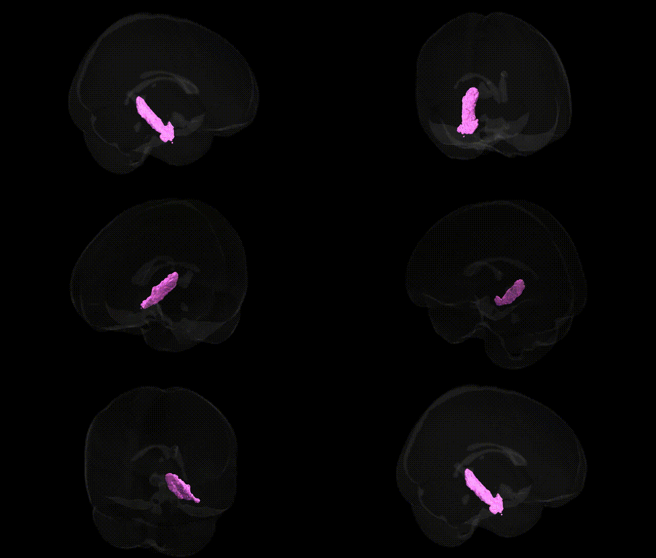
The bilateral GM Hippocampus subiculum L, as defined in the Juelich maxprob thr25 1mm atlas, corresponds to the left subiculum, a key transitional cortical region forming the principal output zone of the hippocampal formation. Located between the CA1 field of the hippocampus proper and the entorhinal and parahippocampal cortices, the subiculum is composed of densely packed pyramidal neurons and displays a characteristic laminar organization that bridges allocortex and neocortex. It plays a central role in relaying processed hippocampal information to widespread cortical and subcortical targets, including the prefrontal cortex, nucleus accumbens, amygdala, and hypothalamus, thereby contributing to episodic memory consolidation, spatial navigation, contextual processing, and stress-related responses. Functionally, the left subiculum is often implicated in verbal and declarative memory networks, and its structural and functional integrity is altered in conditions such as temporal lobe epilepsy, major depressive disorder, and neurodegenerative diseases involving the medial temporal lobe. There is no direct Wikipedia article for this exact atlas label; a closely related structure is the Subiculum.
Genetic associations implicating the hippocampal subiculum, including the left GM Hippocampus subiculum from the Juelich maxprob atlas, largely emerge from imaging genetics and GWAS of hippocampal volume, shape, and connectivity rather than this specific subfield alone. Variants in genes involved in synaptic plasticity and neurodevelopment—such as BDNF (e.g., Val66Met), SLC6A4, and COMT—as well as loci identified in large hippocampal volume GWAS (including regions near ASTN2, DPP4, HRK, and MSRB3) have been associated with structural differences encompassing the subiculum. Polygenic risk scores for Alzheimer’s disease, schizophrenia, and major depressive disorder correlate with hippocampal atrophy and microstructural alterations that prominently involve subicular circuits, and APOE ε4 carrier status is consistently linked to reduced hippocampal integrity and accelerated degeneration affecting subiculum, a key output structure vulnerable in early Alzheimer’s pathology. GWAS of memory performance, cognitive aging, and stress-related traits have connected hippocampal-related genetic variants—including those in FKBP5, NR3C1, and other stress-response genes—to functional changes in subiculum-dependent memory and emotional regulation, while neurodevelopmental risk alleles for disorders such as schizophrenia and bipolar disorder show associations with altered hippocampal subfield morphology in high-resolution MRI studies. Overall, genetic influences on neurodegeneration, synaptic transmission, stress responsivity, and neurodevelopment converge on the hippocampal formation, with the subiculum acting as a critical hub through which these risk variants manifest structural and functional brain phenotypes observed across multiple GWAS and imaging-genetics cohorts.
Overview generated by GPT-4o (2026).
Region ID: 25
Hemisphere: bilateral
Atlas: Juelich maxprob thr25 1mm
Full Quality Version: Download MP4
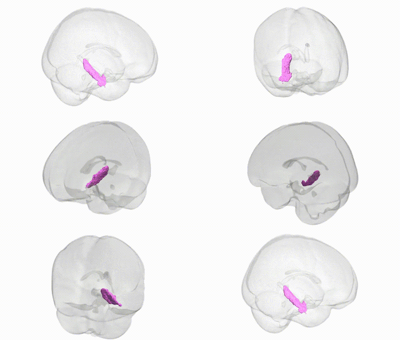
Full Quality Version: Download MP4
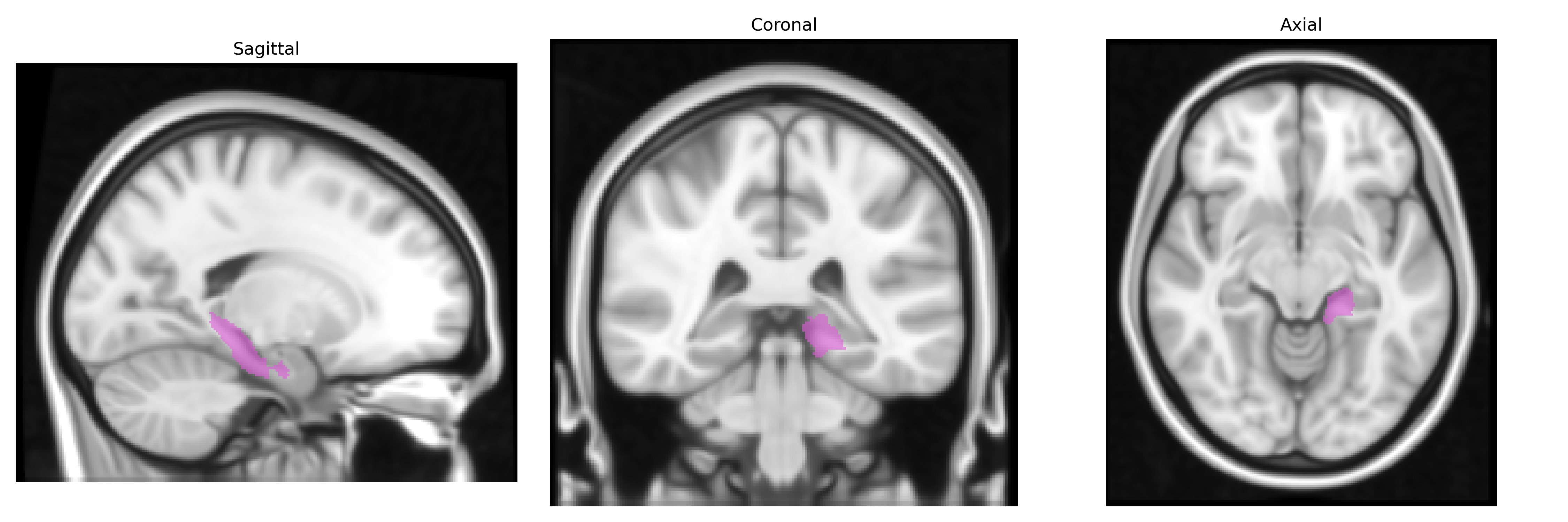
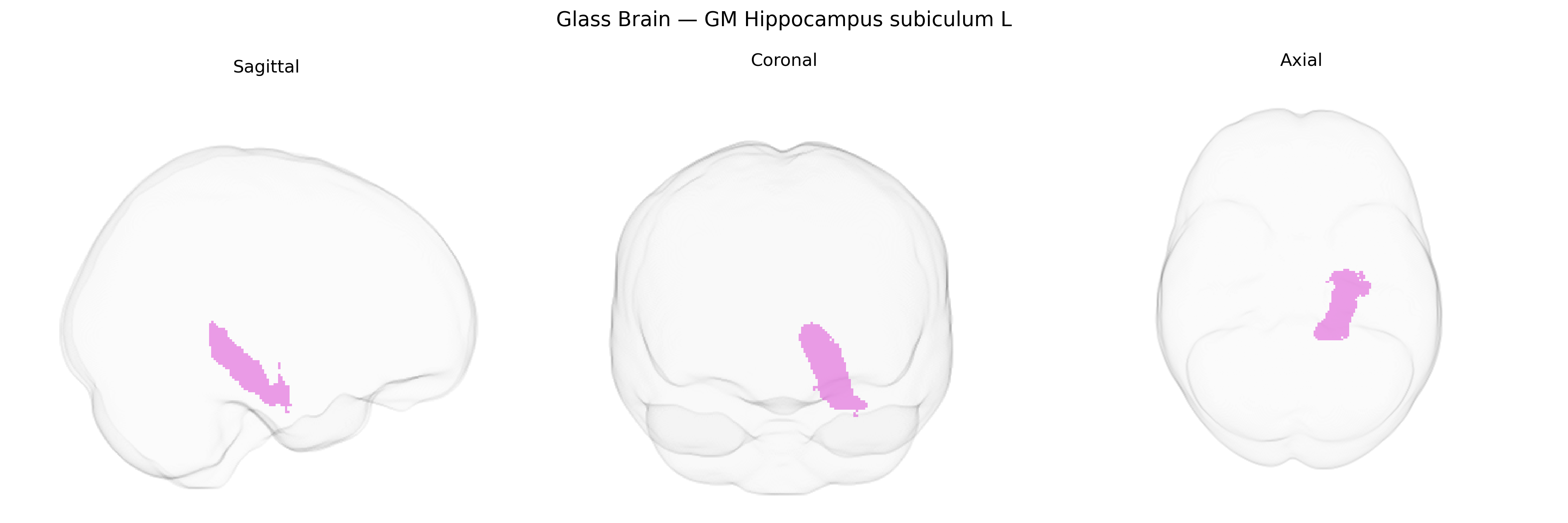
Wali Sidiqyar*, Gaurav Rudravaram*, Elyssa M. McMaster, Trent M. Schwartz, Adam M. Saunders, Kurt G. Schilling, Bennett A. Landman "Introducing SPINS: A Shared Public Visualization Library of Neuroanatomical Structures." Medical Imaging with Deep Learning- short paper
This resource is licensed under CC0 1.0 Universal (Public Domain).