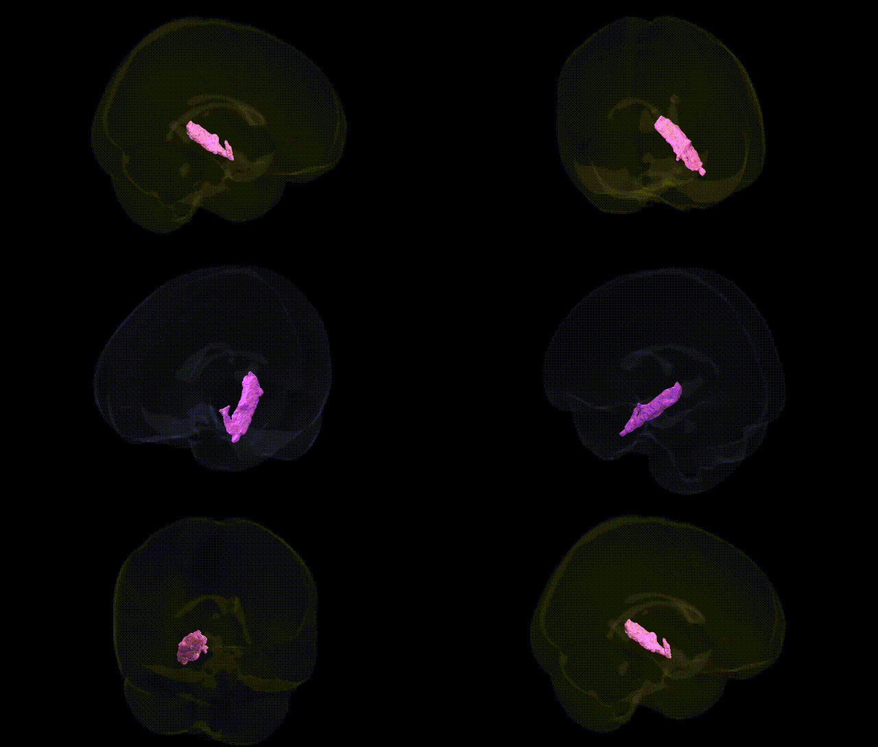
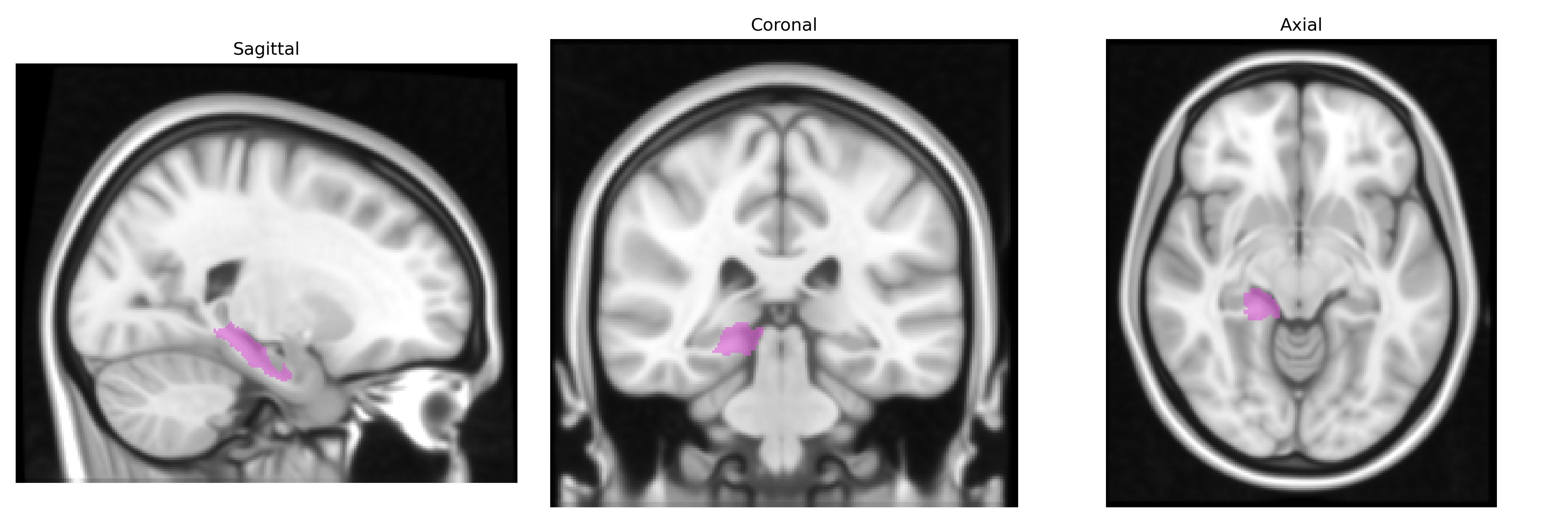
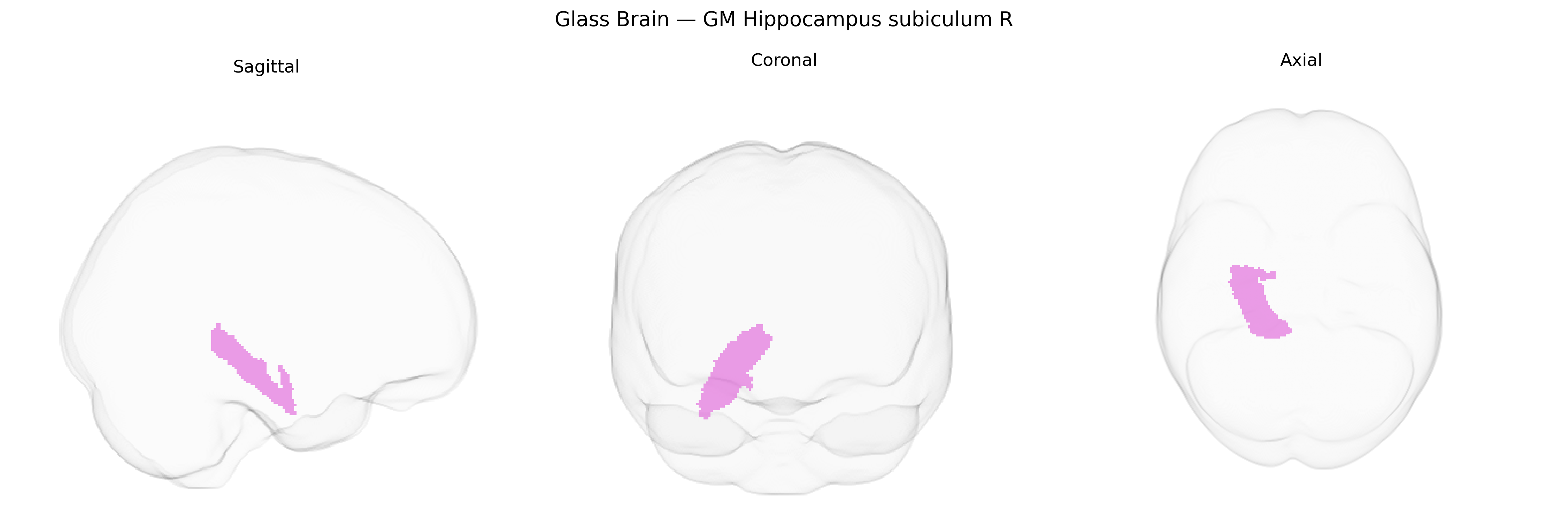
The bilateral GM Hippocampus subiculum R, as defined in the Juelich maxprob thr25 1 mm atlas, corresponds to the right subicular portion of the hippocampal formation, a transitional cortical region positioned between the CA1 field of the hippocampus proper and the entorhinal cortex. It serves as a major output node of the hippocampus, receiving processed information from CA1 and projecting extensively to limbic and associative structures, including the entorhinal cortex, amygdala, nucleus accumbens, mammillary bodies, and prefrontal cortices. Cytoarchitectonically, the subiculum exhibits a laminar organization intermediate between archicortex and neocortex, reflecting its role as a gateway for hippocampal signals involved in spatial navigation, memory consolidation, and contextual representation. There is no direct Wikipedia article for “Hippocampus subiculum R”; a related structure is the Subiculum.
The hippocampal subiculum, as defined in the Juelich maxprob thr25 1 mm atlas, has been implicated in genetic studies primarily through its role in hippocampal structure, memory, and stress-related circuitry, with findings generally bilateral rather than strictly right-lateralized. GWAS of hippocampal volume and subfields, including the subiculum, have identified common variants in and around genes such as SLC4A10, DPP4, ASTN2, MSRB3, and those within the 12q24 and 2q24 regions, which influence hippocampal morphology and are often shared across multiple limbic structures. Polygenic risk scores for Alzheimer’s disease, schizophrenia, major depressive disorder, and bipolar disorder show associations with reduced hippocampal and subicular volumes, consistent with the subiculum’s involvement in memory impairment, psychosis-related circuit dysfunction, and mood regulation. Variants in APOE (particularly ε4), BIN1, and CLU, strongly implicated in Alzheimer’s disease GWAS, are associated with atrophy in hippocampal regions that include the subiculum, while schizophrenia and depression GWAS implicate synaptic and glutamatergic genes (e.g., GRM3, CACNA1C, GRIN2A) that map onto networks in which the subiculum is a key output structure. Imaging–genetics studies further link hippocampal subiculum measures to genetic risk for PTSD, anxiety traits, and cognitive performance, indicating that the genetic architecture governing hippocampal subiculum structure and function overlaps substantially with that for neurodegenerative and major psychiatric disorders as well as normal variation in memory and general cognitive ability.
Overview generated by GPT-4o (2026).
Region ID: 26
Hemisphere: bilateral
Atlas: Juelich maxprob thr25 1mm
Full Quality Version: Download MP4
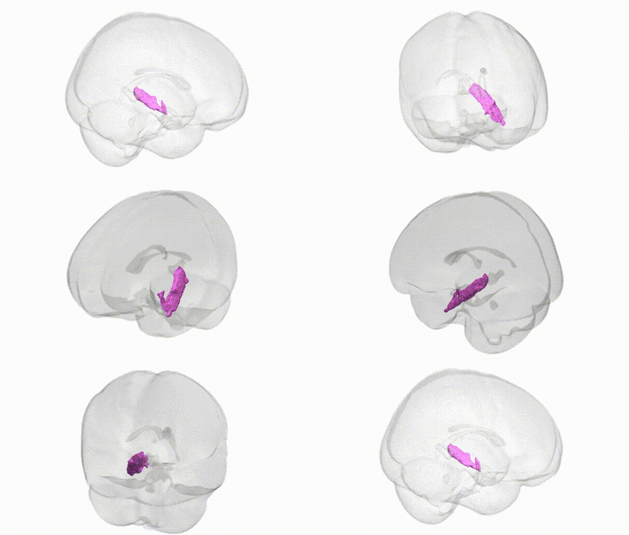
Full Quality Version: Download MP4
Wali Sidiqyar*, Gaurav Rudravaram*, Elyssa M. McMaster, Trent M. Schwartz, Adam M. Saunders, Kurt G. Schilling, Bennett A. Landman "Introducing SPINS: A Shared Public Visualization Library of Neuroanatomical Structures." Medical Imaging with Deep Learning- short paper
This resource is licensed under CC0 1.0 Universal (Public Domain).