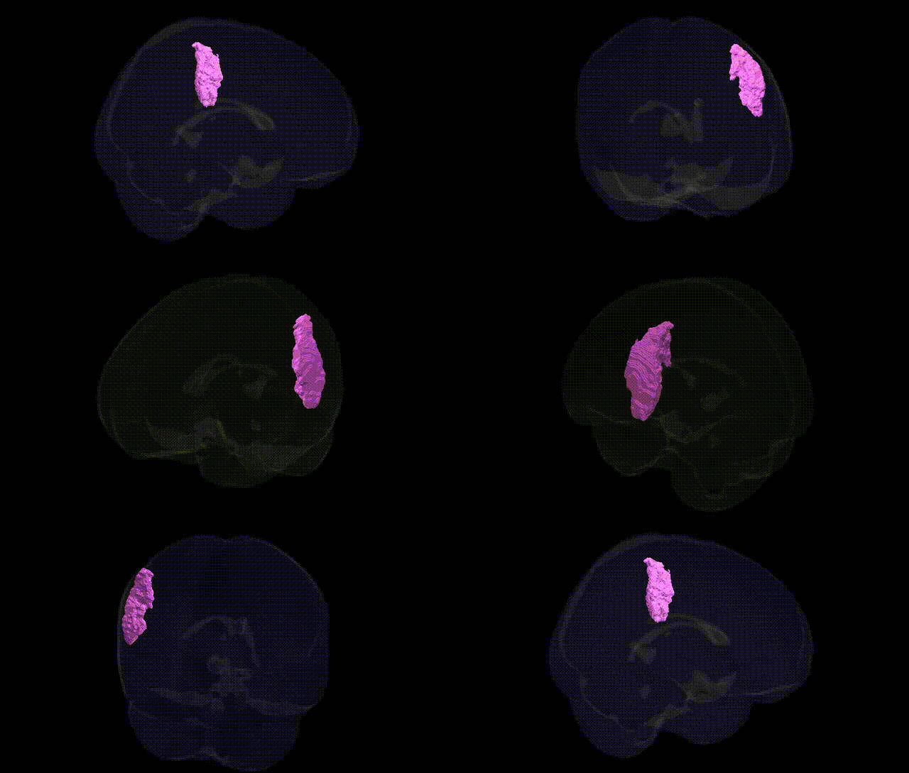
The bilateral GM Inferior parietal lobule PF R (Juelich maxprob thr25 1mm Atlas) corresponds to a cytoarchitectonic subregion within the inferior parietal lobule (IPL), associated primarily with multimodal integration, higher-order somatosensory processing, and aspects of attention and social cognition. Located in the posterior parietal cortex, this PF subdivision forms part of the supramarginal gyrus territory and participates in networks supporting action observation, tool use, language-related processes (particularly phonological and semantic integration), and allocation of spatial attention. Functionally, it interfaces with frontal premotor and prefrontal areas as well as temporal regions, contributing to sensorimotor transformation, working memory, and perspective-taking. There is no direct Wikipedia article for “PF R”; a closely related structure is the Inferior parietal lobule.
The bilateral GM Inferior parietal lobule PF R region, a higher-order multimodal association area implicated in language, attention, social cognition, and tool use, has been indirectly associated with a range of genetic findings from neuroimaging and disorder GWAS, though few studies target the Juelich PF subregion specifically. Imaging genetics research has linked common variants in genes such as CNTNAP2, FOXP2, DCDC2, KIAA0319, and ROBO1 (involved in language and reading/dyslexia risk) to structural and functional variation in inferior parietal and supramarginal areas that overlap PF territories. Large neuroimaging GWAS consortia (e.g., ENIGMA, UK Biobank) have identified polygenic contributions to parietal cortical thickness and surface area, implicating loci in neurodevelopmental and synaptic genes (including MAPT region haplotypes, cytoskeletal and cell-adhesion genes), with parietal lobule morphometry showing significant genetic correlation with general cognitive ability, educational attainment, and reading-related traits. Disorders linked to genetic variation affecting inferior parietal regions include developmental dyslexia, autism spectrum disorder, schizophrenia, and attention-deficit/hyperactivity disorder, where risk loci in synaptic and neurodevelopmental genes (e.g., GRIN2A, NRXN1, SHANK3, and common polygenic risk scores) have been associated with altered activation or connectivity in PF-like parietal areas during language, working memory, and social cognition tasks. Additionally, parietal association cortex, including PF, shows heritable patterns of connectivity with fronto-temporal language and default-mode networks, with GWAS of resting-state network topology indicating shared genetic influences between parietal hub architecture and traits such as intelligence, mathematical ability, and risk for major depressive disorder, although these findings generally map to broader inferior parietal territories rather than the PF R parcel alone.
Overview generated by GPT-4o (2026).
Region ID: 28
Hemisphere: bilateral
Atlas: Juelich maxprob thr25 1mm
Full Quality Version: Download MP4
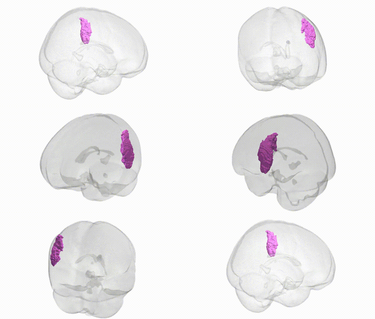
Full Quality Version: Download MP4
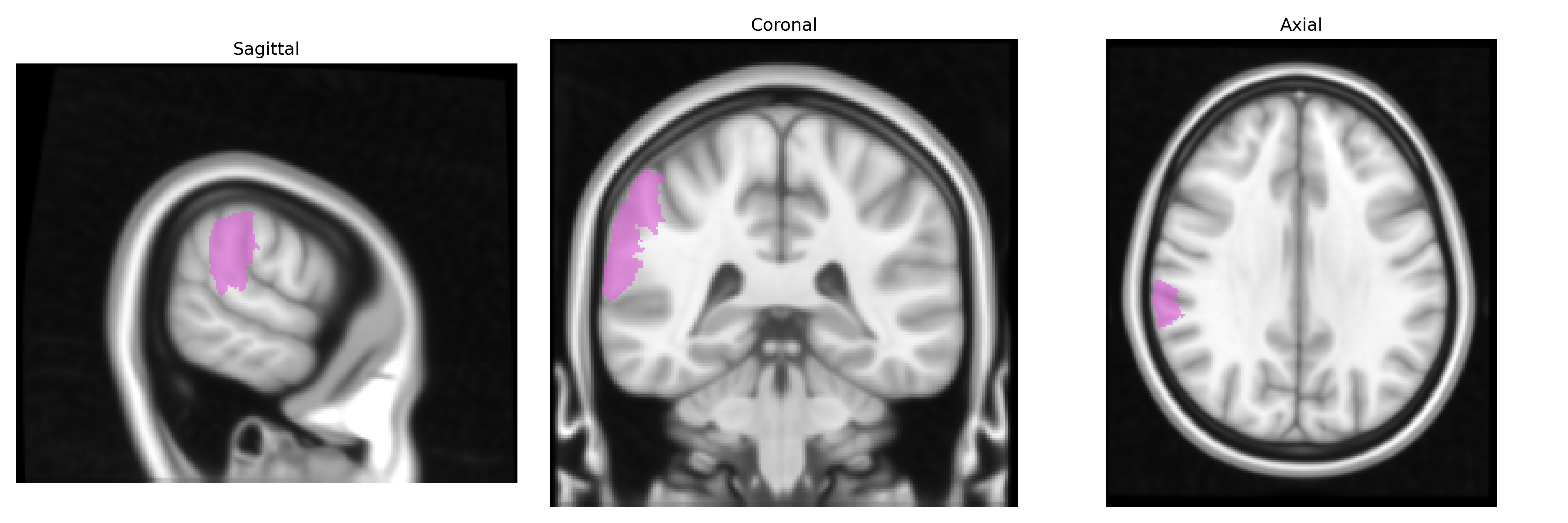
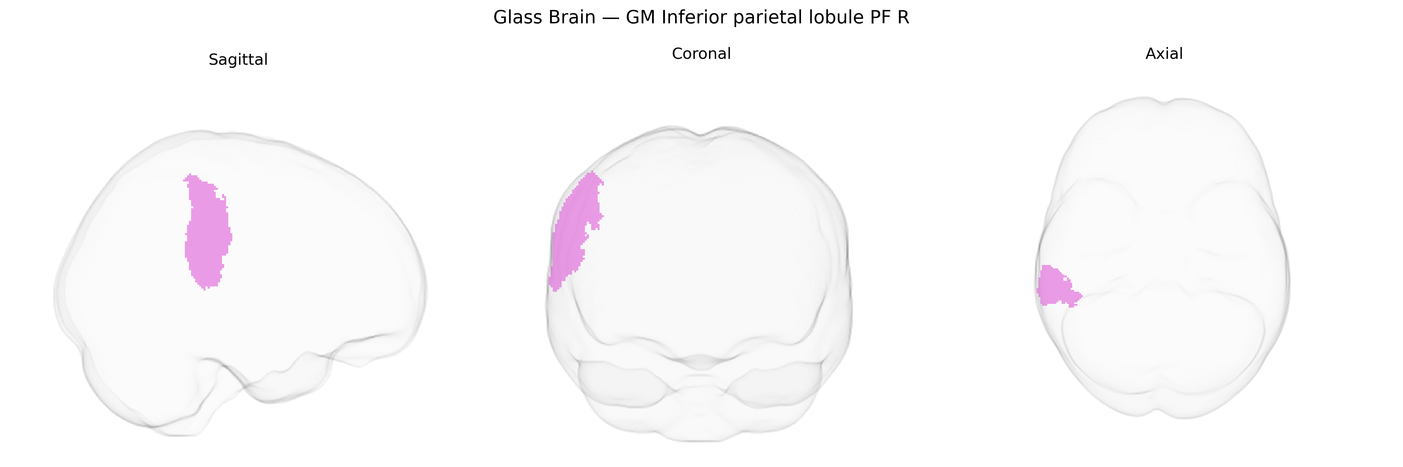
Wali Sidiqyar*, Gaurav Rudravaram*, Elyssa M. McMaster, Trent M. Schwartz, Adam M. Saunders, Kurt G. Schilling, Bennett A. Landman "Introducing SPINS: A Shared Public Visualization Library of Neuroanatomical Structures." Medical Imaging with Deep Learning- short paper
This resource is licensed under CC0 1.0 Universal (Public Domain).