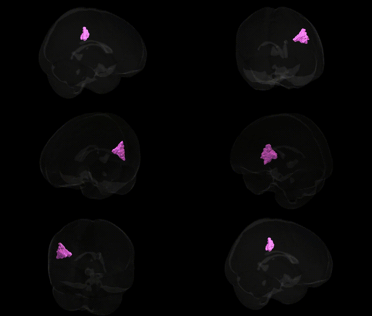
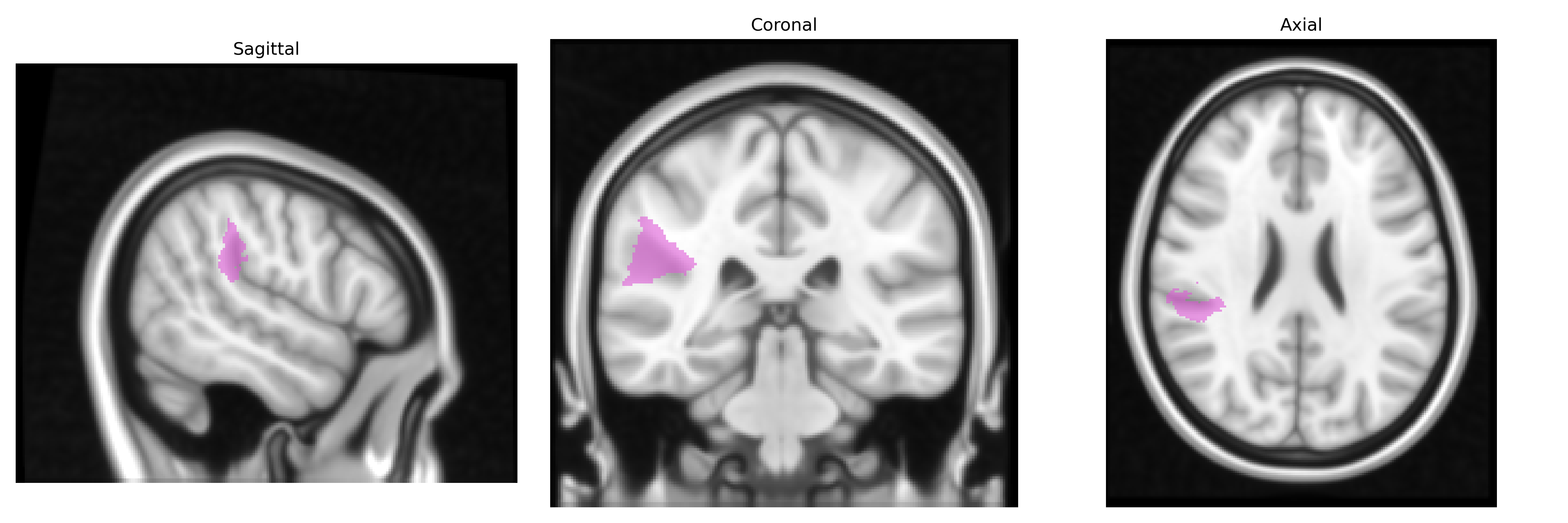
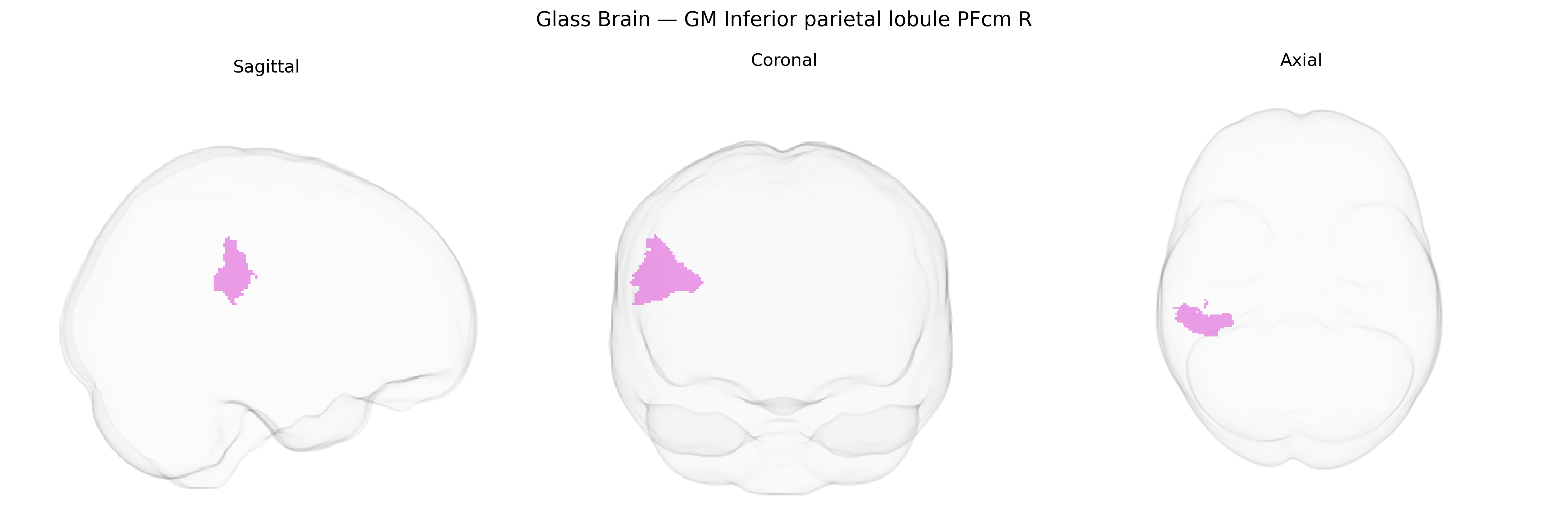
The bilateral GM Inferior parietal lobule PFcm R (from the Juelich maxprob thr25 1mm atlas) refers to a cytoarchitectonically defined subregion (PFcm) within the inferior parietal lobule, located in the posterior parietal cortex and associated with multimodal integration of visual, auditory, and somatosensory information. This region is implicated in higher-order cognitive functions such as attention reorientation, action understanding, and aspects of social cognition, and likely participates in fronto-parietal networks supporting working memory and sensorimotor transformation. PFcm is part of the broader inferior parietal lobule complex and corresponds to a subdivision of the supramarginal/angular gyrus region, although it does not have a dedicated Wikipedia entry; for general reference, see the related structure Inferior parietal lobule.
The bilateral inferior parietal lobule, including the PFcm area on the right as defined in the Juelich maxprob thr25 1 mm atlas, has been implicated in multiple genetic and genome-wide association studies through its roles in higher-order cognition, language, attention, and social processing, although PFcm-specific genetic associations remain indirect and regionally coarse. GWAS and imaging-genetics studies have linked variation in parietal cortical thickness, surface area, and functional connectivity to loci involved in neurodevelopmental pathways (for example, genes affecting synaptic formation, axon guidance, and cortical patterning such as BDNF, DISC1, and FOXP2-related networks), and have associated inferior parietal morphology or activation with risk for schizophrenia, autism spectrum disorder, attention-deficit/hyperactivity disorder, major depressive disorder, and Alzheimer’s disease, as well as with cognitive traits including general intelligence, working memory, reading and language abilities, and educational attainment. Large-scale consortia such as ENIGMA and UK Biobank have reported heritable variation in inferior parietal structure and function and identified polygenic influences overlapping with neurotransmission (e.g., glutamatergic and GABAergic signaling), synaptic plasticity, and immune-related genes, which may contribute to vulnerability for psychiatric and neurodegenerative conditions involving parietal dysfunction. However, current genetic evidence typically targets broader inferior parietal or supramarginal/angular gyrus territories rather than the PFcm subregion specifically, so associations with PFcm R remain inferred from these larger parietal networks rather than directly resolved at the fine-grained cytoarchitectonic level.
Overview generated by GPT-4o (2026).
Region ID: 30
Hemisphere: bilateral
Atlas: Juelich maxprob thr25 1mm
Full Quality Version: Download MP4
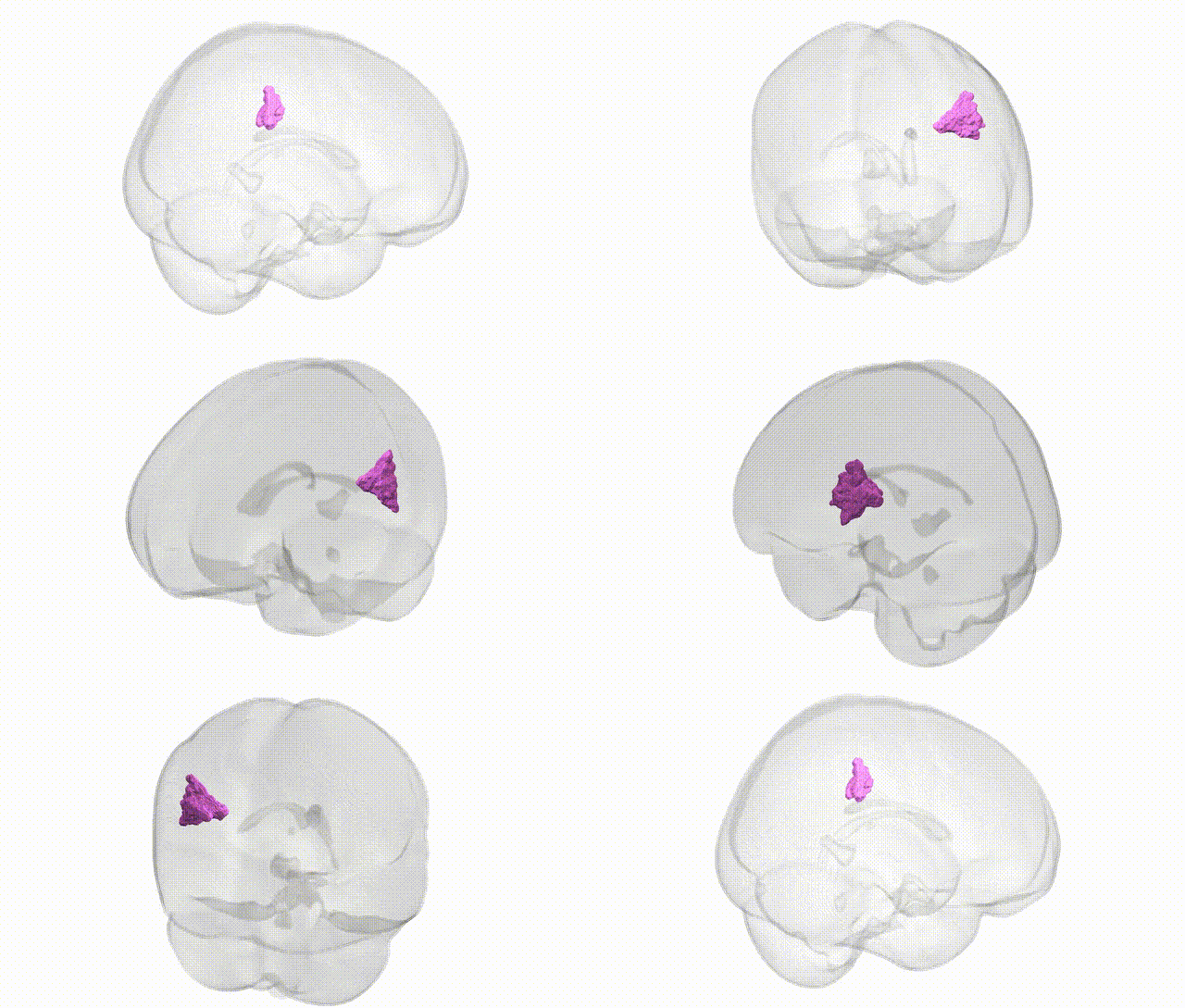
Full Quality Version: Download MP4
Wali Sidiqyar*, Gaurav Rudravaram*, Elyssa M. McMaster, Trent M. Schwartz, Adam M. Saunders, Kurt G. Schilling, Bennett A. Landman "Introducing SPINS: A Shared Public Visualization Library of Neuroanatomical Structures." Medical Imaging with Deep Learning- short paper
This resource is licensed under CC0 1.0 Universal (Public Domain).