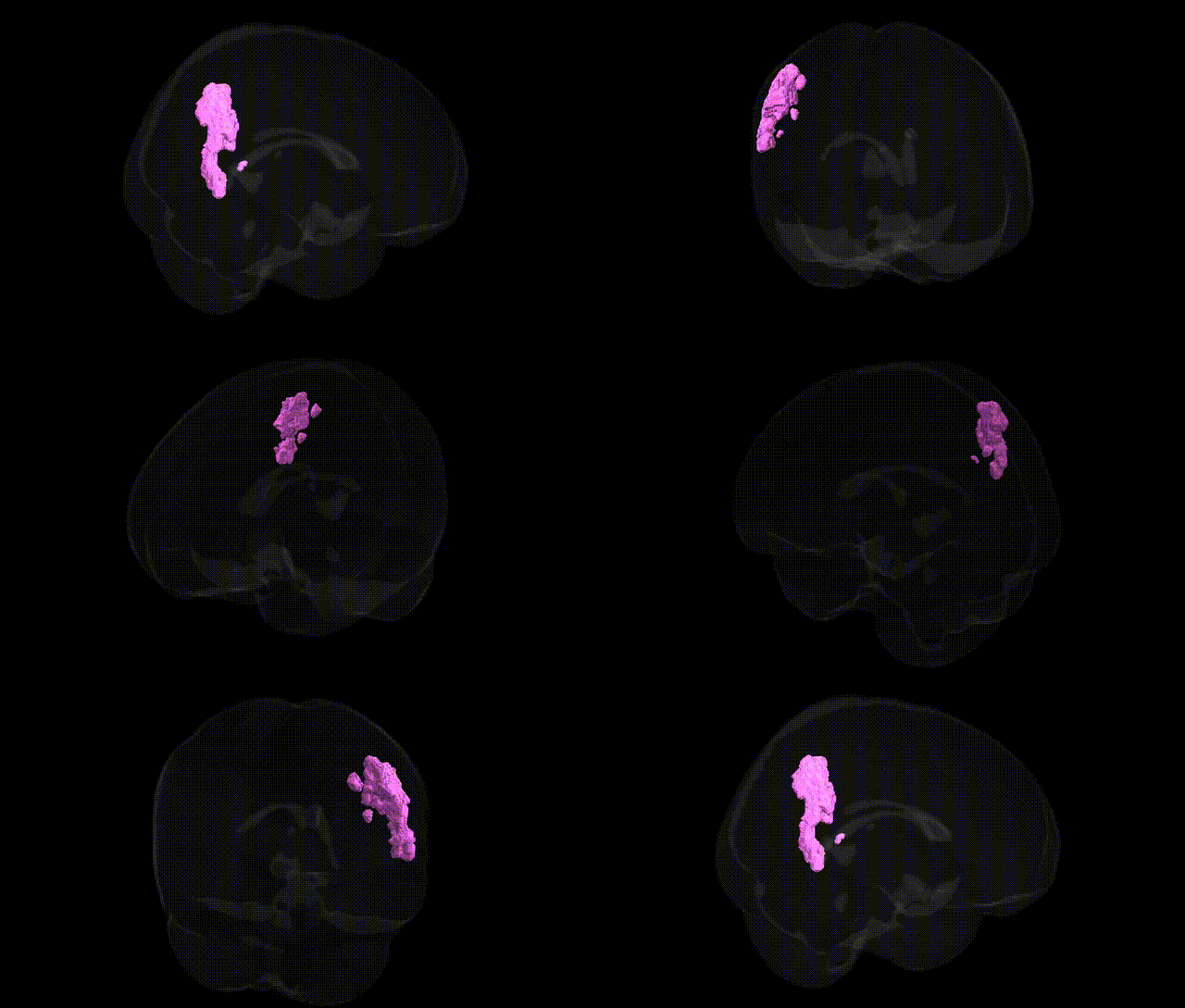
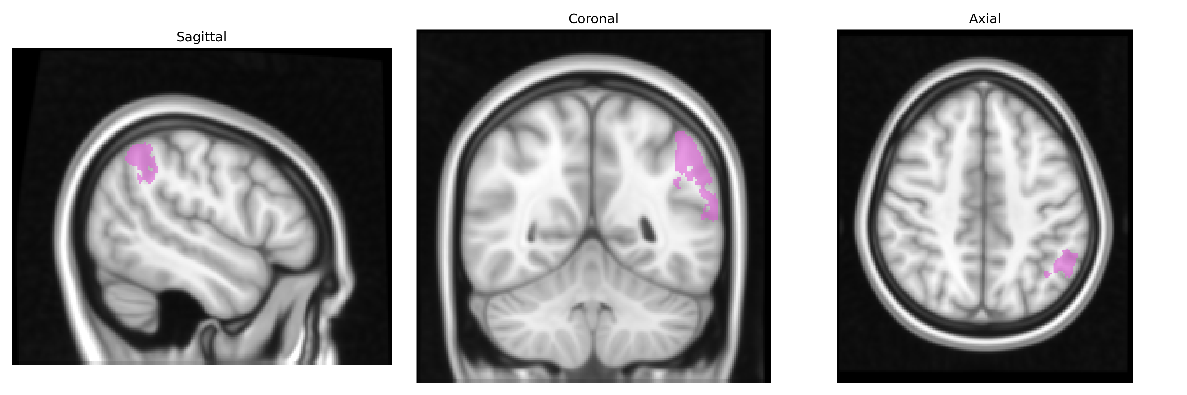
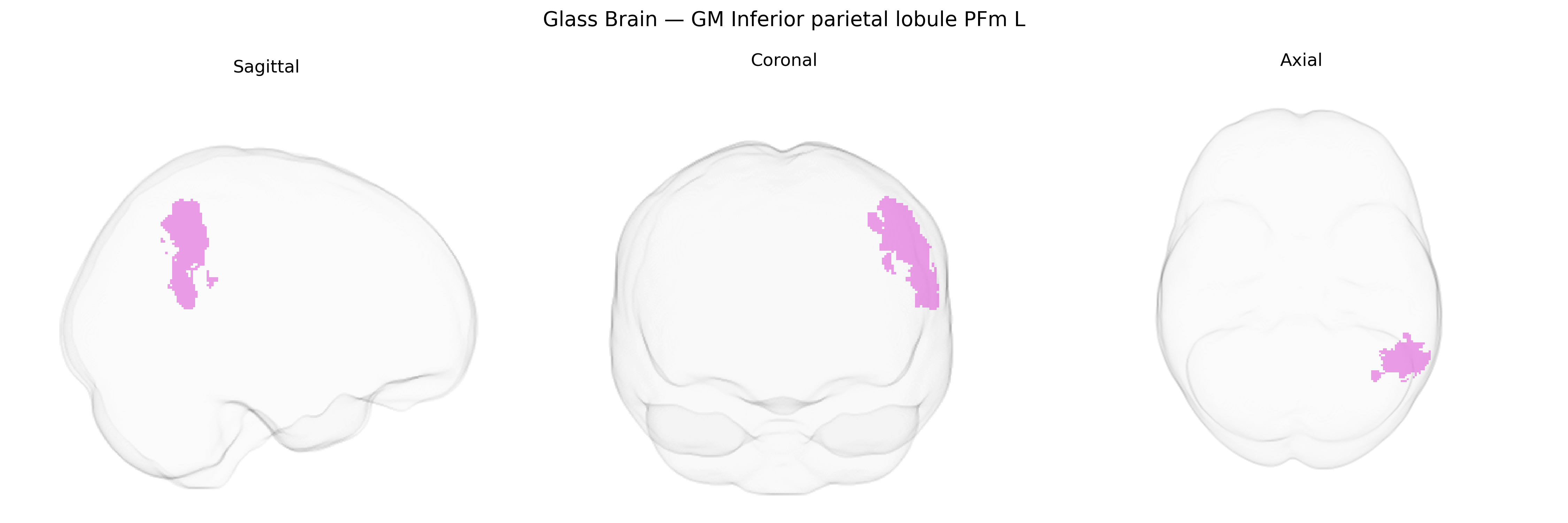
The bilateral GM Inferior parietal lobule PFm L, as defined in the Juelich maxprob thr25 1mm Atlas, corresponds to a cytoarchitectonic subregion (PFm) within the inferior parietal lobule of the left hemisphere, associated predominantly with higher-order multimodal integration and aspects of social cognition. This region participates in processing and integrating visual, somatosensory, and auditory information, and is implicated in functions such as action observation, tool use, spatial attention, and aspects of language and conceptual understanding. PFm lies within the broader inferior parietal cortex, which forms part of the posterior parietal network involved in praxis, body-schema representation, and the interface between perception and action. There is no direct Wikipedia article for PFm; a closely related and encompassing structure is the inferior parietal lobule: Inferior parietal lobule.
The bilateral GM Inferior parietal lobule PFm L region (Juelich maxprob thr25 1mm) lies within the inferior parietal cortex, a hub repeatedly implicated in polygenic influences on higher cognition, social processing, and neuropsychiatric risk. GWAS of cortical thickness, surface area, and regional volume have identified common variants in and near genes such as microtubule-associated protein tau (MAPT), WNT pathway genes, and synaptic/neuronal development loci that modulate parietal morphology, including inferior parietal subregions overlapping PFm. Large-scale imaging–genetics consortia (e.g., ENIGMA, UK Biobank) report that parietal measures show significant SNP-based heritability and share genetic correlations with general intelligence, educational attainment, reading ability, and numerical cognition, as well as with attention-deficit/hyperactivity disorder, autism spectrum disorder, schizophrenia, and major depression, all disorders in which altered inferior parietal structure or function is reported. Alzheimer’s disease GWAS loci (e.g., APOE, CLU, PICALM, BIN1) and polygenic risk scores also show associations with atrophy patterns and connectivity changes encompassing inferior parietal regions, including PFm, consistent with the role of this area in default mode and temporoparietal networks vulnerable in prodromal and clinical neurodegeneration. Collectively, genetic studies indicate that PFm-related parietal variation reflects a distributed polygenic architecture shared with cognitive ability, psychiatric and neurodevelopmental disorders, and dementia risk, rather than a single locus specifically tied to this cytoarchitectonic field.
Overview generated by GPT-4o (2026).
Region ID: 31
Hemisphere: bilateral
Atlas: Juelich maxprob thr25 1mm
Full Quality Version: Download MP4
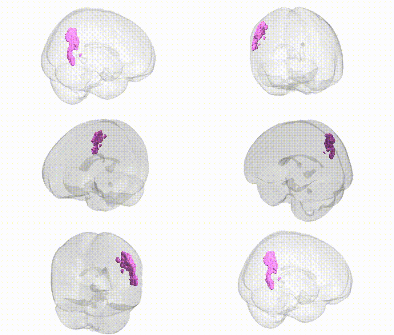
Full Quality Version: Download MP4
Wali Sidiqyar*, Gaurav Rudravaram*, Elyssa M. McMaster, Trent M. Schwartz, Adam M. Saunders, Kurt G. Schilling, Bennett A. Landman "Introducing SPINS: A Shared Public Visualization Library of Neuroanatomical Structures." Medical Imaging with Deep Learning- short paper
This resource is licensed under CC0 1.0 Universal (Public Domain).