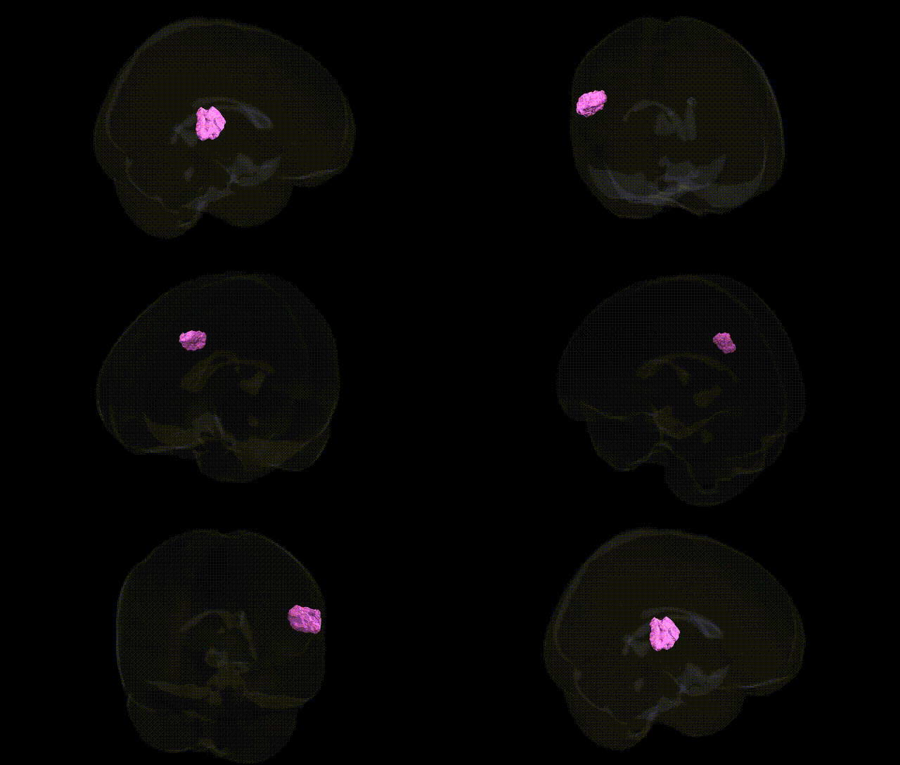
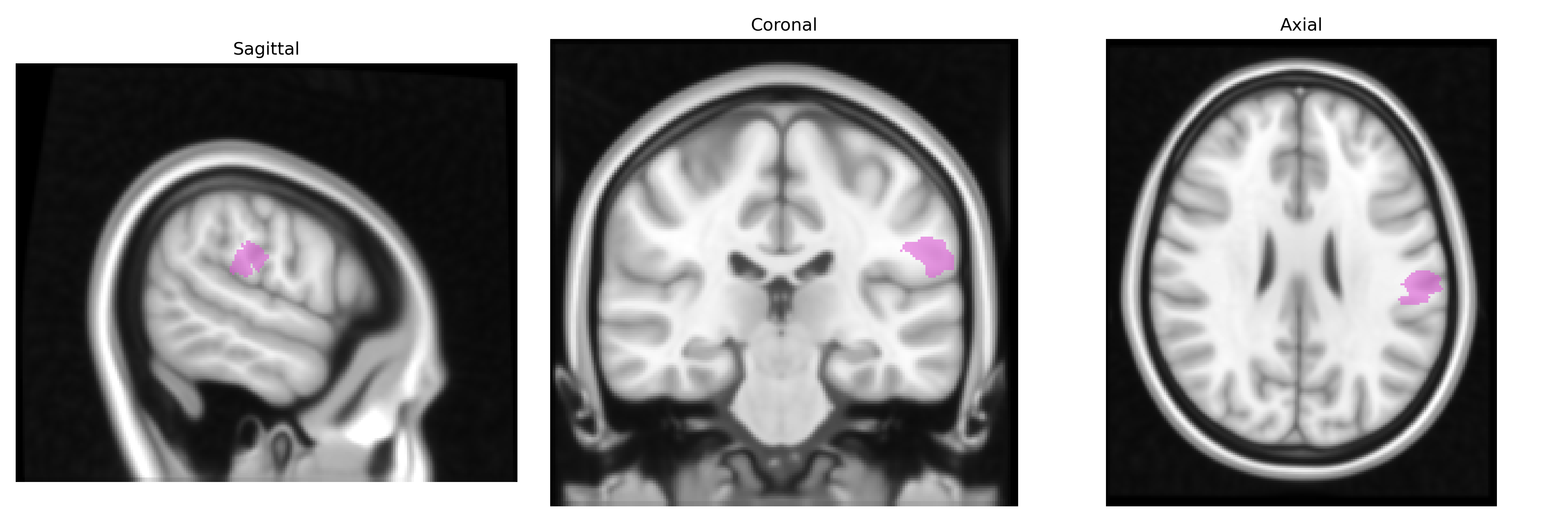
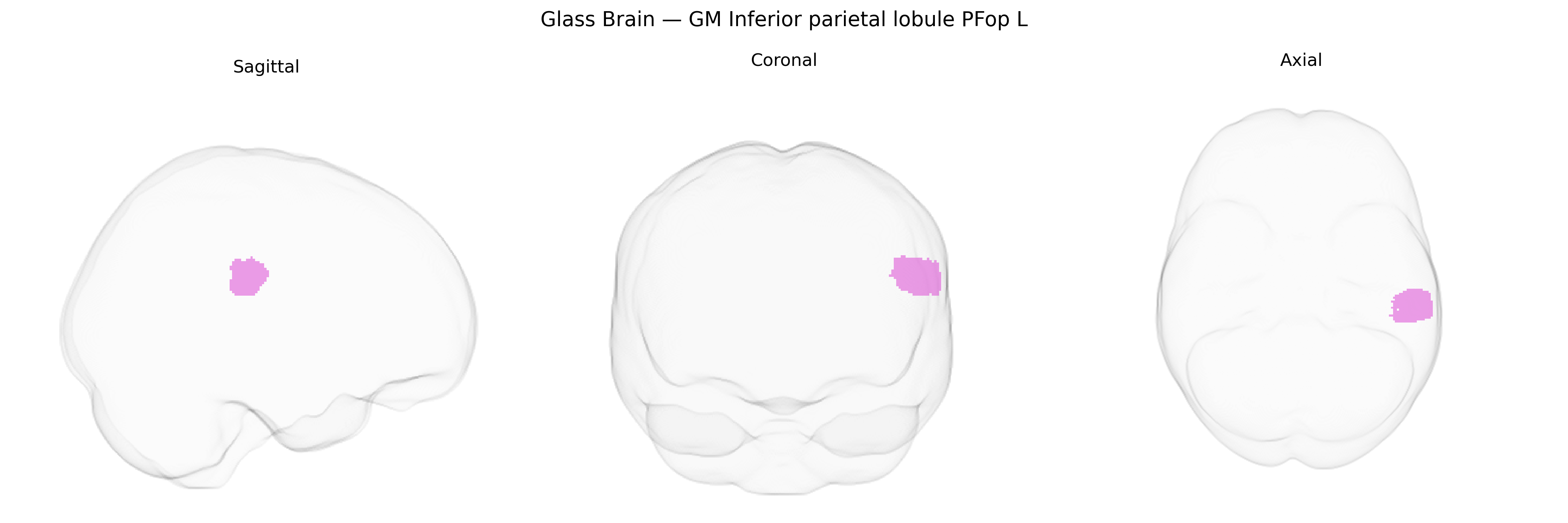
The bilateral GM Inferior parietal lobule PFop L (Juelich maxprob thr25 1mm Atlas) corresponds to a cytoarchitectonic subregion of the inferior parietal lobule, located in the posterior parietal cortex at the junction of the supramarginal and superior temporal regions, and is considered part of the parietal operculum and perisylvian association cortex. This region is implicated in multimodal integration of somatosensory, auditory, and visual information, and contributes to higher-order functions such as language processing (particularly phonological and articulatory aspects), praxis, attention reorienting, and aspects of social cognition. Neuronal populations in PFop exhibit dense connections with frontal premotor and inferior frontal language regions, as well as temporal association cortices, supporting roles in sensorimotor transformation for speech, action observation and imitation, and the integration of contextual information into perceptual and conceptual representations. There is no direct Wikipedia article for PFop; a related area is the Inferior parietal lobule.
The bilateral GM Inferior parietal lobule PFop L region (Juelich maxprob thr25 1mm) corresponds roughly to part of the inferior parietal cortex involved in language, praxis, and multimodal integration, and its genetic associations have been inferred primarily from imaging–genetics and GWAS-based imaging studies rather than region-specific candidate-gene work. Large-scale GWAS of cortical thickness and surface area (e.g., ENIGMA, UK Biobank) have identified loci in or near genes such as MIR924HG, LINC01158, LPAR1, and DAAM1 that influence parietal lobe morphology, including inferior parietal regions; polygenic scores related to general cognitive ability, educational attainment (e.g., loci near MAPT, KIAA0319, and other neurodevelopmental genes), and schizophrenia have shown associations with structure and function in parietal cortices that encompass PFop territories. Genetic variants tied to language-related disorders (such as dyslexia-associated genes including DCDC2, KIAA0319, and ROBO1) and autism spectrum conditions have been linked to structural or functional alterations in inferior parietal regions, while risk loci for Alzheimer’s disease (e.g., APOE ε4 and nearby modulators of amyloid and tau processing) have been associated with altered parietal atrophy patterns and network connectivity that include PFop-adjacent cortex. Additionally, genome-wide polygenic risk for attention-deficit/hyperactivity disorder and major depressive disorder has been associated with differences in parietal functional networks and default mode connectivity that involve inferior parietal lobule subregions, though these effects are typically distributed and not specific to PFop. Overall, genetic findings implicate a broad set of neurodevelopmental, synaptic, and plasticity-related genes in shaping structure and function of the inferior parietal cortex, with PFop L likely participating in these polygenic influences on cognition, language, psychiatric vulnerability, and neurodegenerative risk, even though direct PFop-specific GWAS signals are not yet well delineated.
Overview generated by GPT-4o (2026).
Region ID: 33
Hemisphere: bilateral
Atlas: Juelich maxprob thr25 1mm
Full Quality Version: Download MP4
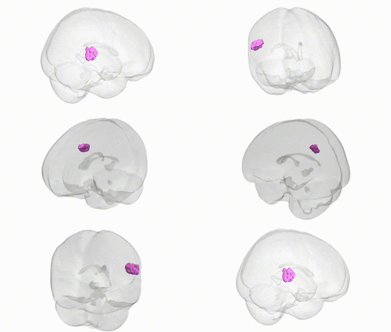
Full Quality Version: Download MP4
Wali Sidiqyar*, Gaurav Rudravaram*, Elyssa M. McMaster, Trent M. Schwartz, Adam M. Saunders, Kurt G. Schilling, Bennett A. Landman "Introducing SPINS: A Shared Public Visualization Library of Neuroanatomical Structures." Medical Imaging with Deep Learning- short paper
This resource is licensed under CC0 1.0 Universal (Public Domain).