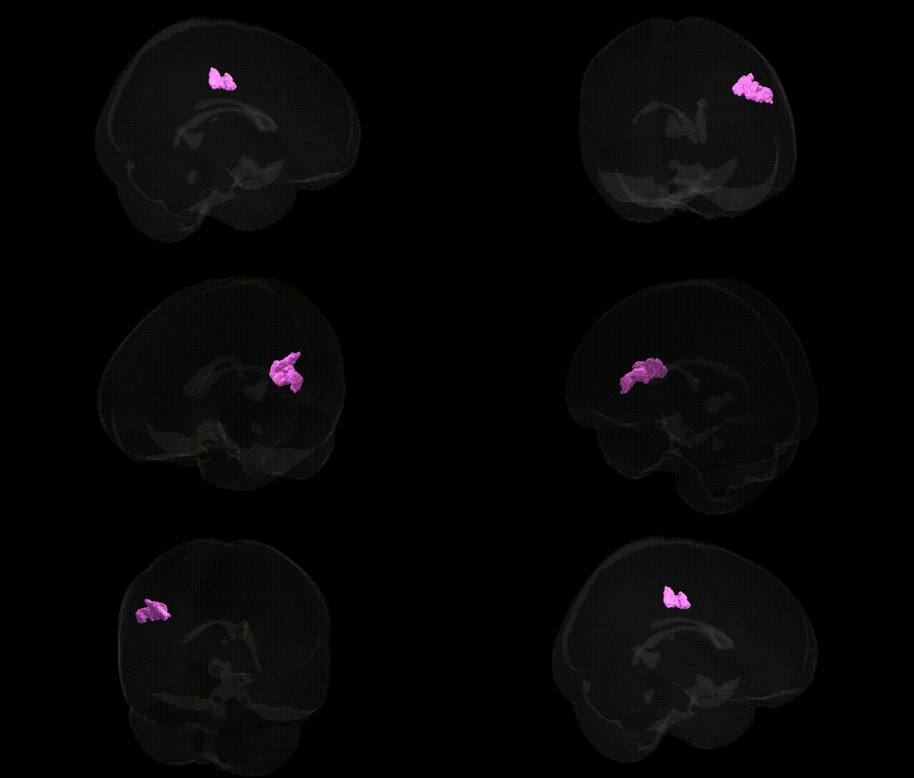
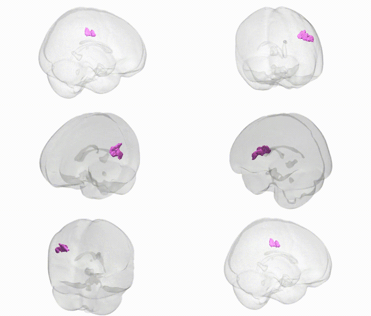
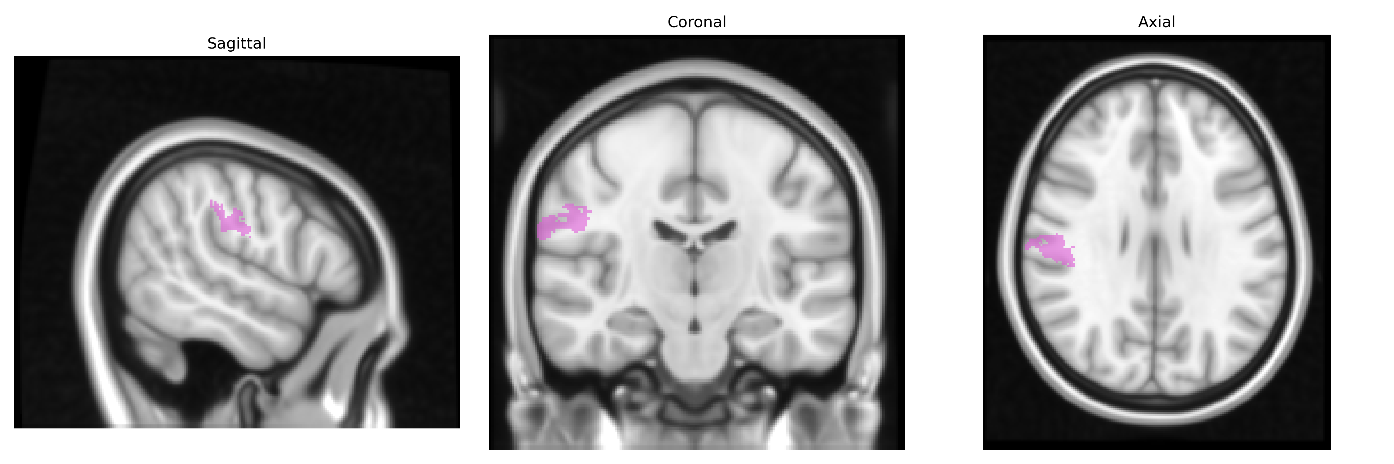
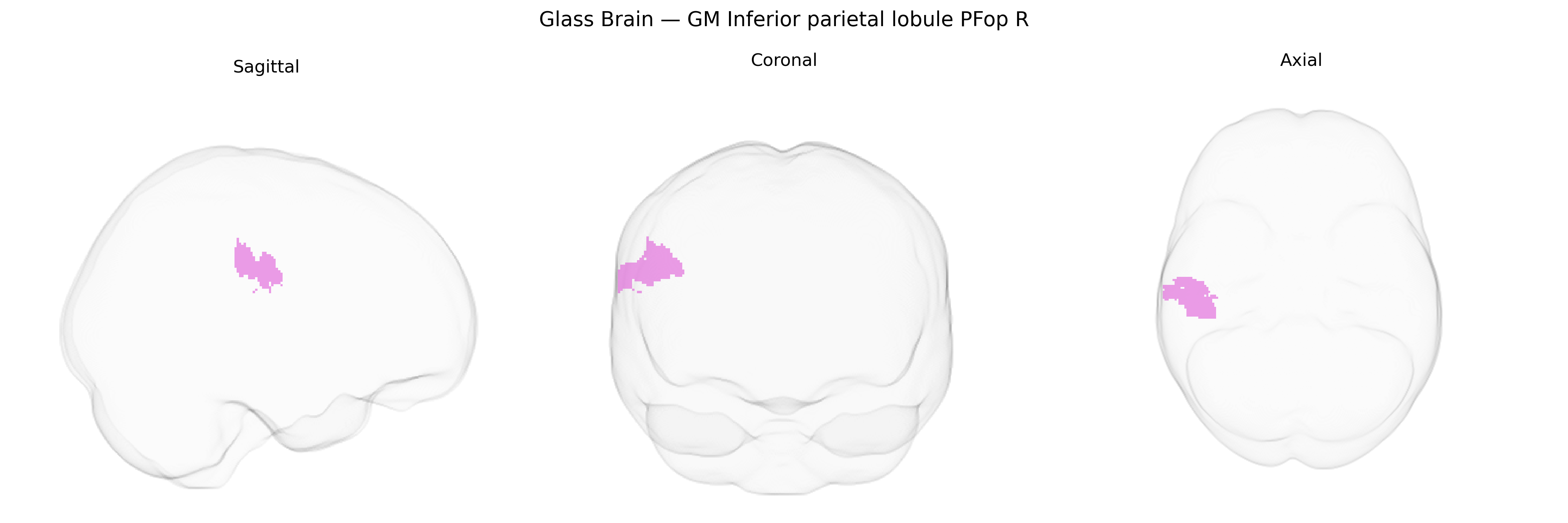
The bilateral GM Inferior parietal lobule PFop R, as defined in the Juelich maxprob thr25 1mm Atlas, corresponds to an opercular subdivision (PFop) of the inferior parietal lobule in the right hemisphere, encompassing gray matter in the supramarginal gyrus near the parietal–temporal–frontal junction. This cytoarchitectonically defined region is part of the inferior parietal cortex implicated in multimodal sensory integration, higher-order somatosensory processing, and aspects of language and social cognition, including action understanding and phonological processing. PFop is structurally connected with premotor, prefrontal, and temporal cortices, and functionally contributes to networks supporting attention, working memory, and sensorimotor transformations. There is no direct Wikipedia article for PFop; a closely related structure is the supramarginal gyrus: Supramarginal gyrus.
The bilateral inferior parietal lobule PFop (R), as defined in the Juelich maxprob atlas, lies within a parietal region repeatedly implicated in genetic studies of higher cognition, language, attention, and neuropsychiatric risk, though most findings target the broader inferior parietal cortex rather than PFop specifically. GWAS of cortical structure have identified common variants in genes linked to synaptic development and neurogenesis (e.g., variants near MEF2C, DACT1, and other neurodevelopmental loci) that influence parietal cortical thickness and surface area, with inferior parietal measures showing moderate heritability. Parietal association cortex, including PFop, has been associated via imaging-genetics and polygenic risk approaches with schizophrenia, autism spectrum disorder, and attention-deficit/hyperactivity disorder, typically showing altered cortical morphology or connectivity that covaries with polygenic risk scores derived from large psychiatric GWAS consortia. Inferior parietal regions are also frequent readouts in genetic studies of intelligence, educational attainment, and reading/language abilities (e.g., dyslexia), where risk alleles in genes involved in axon guidance and cortical patterning relate to variations in parietal activation and structure during language and working-memory tasks. Additionally, GWAS and imaging-genetics analyses of Alzheimer’s disease and related dementias report genetic influences (for example APOE and other AD-risk loci) on atrophy patterns and functional disruption in inferior parietal areas, which contribute to default mode and multimodal integration networks; however, current evidence localizes these effects at the level of broader parietal lobule parcels rather than the PFop subregion alone, so PFop-specific genetic associations remain indirect and inferred from the larger inferior parietal territory.
Overview generated by GPT-4o (2026).
Region ID: 34
Hemisphere: bilateral
Atlas: Juelich maxprob thr25 1mm
Full Quality Version: Download MP4
Full Quality Version: Download MP4
Wali Sidiqyar*, Gaurav Rudravaram*, Elyssa M. McMaster, Trent M. Schwartz, Adam M. Saunders, Kurt G. Schilling, Bennett A. Landman "Introducing SPINS: A Shared Public Visualization Library of Neuroanatomical Structures." Medical Imaging with Deep Learning- short paper
This resource is licensed under CC0 1.0 Universal (Public Domain).