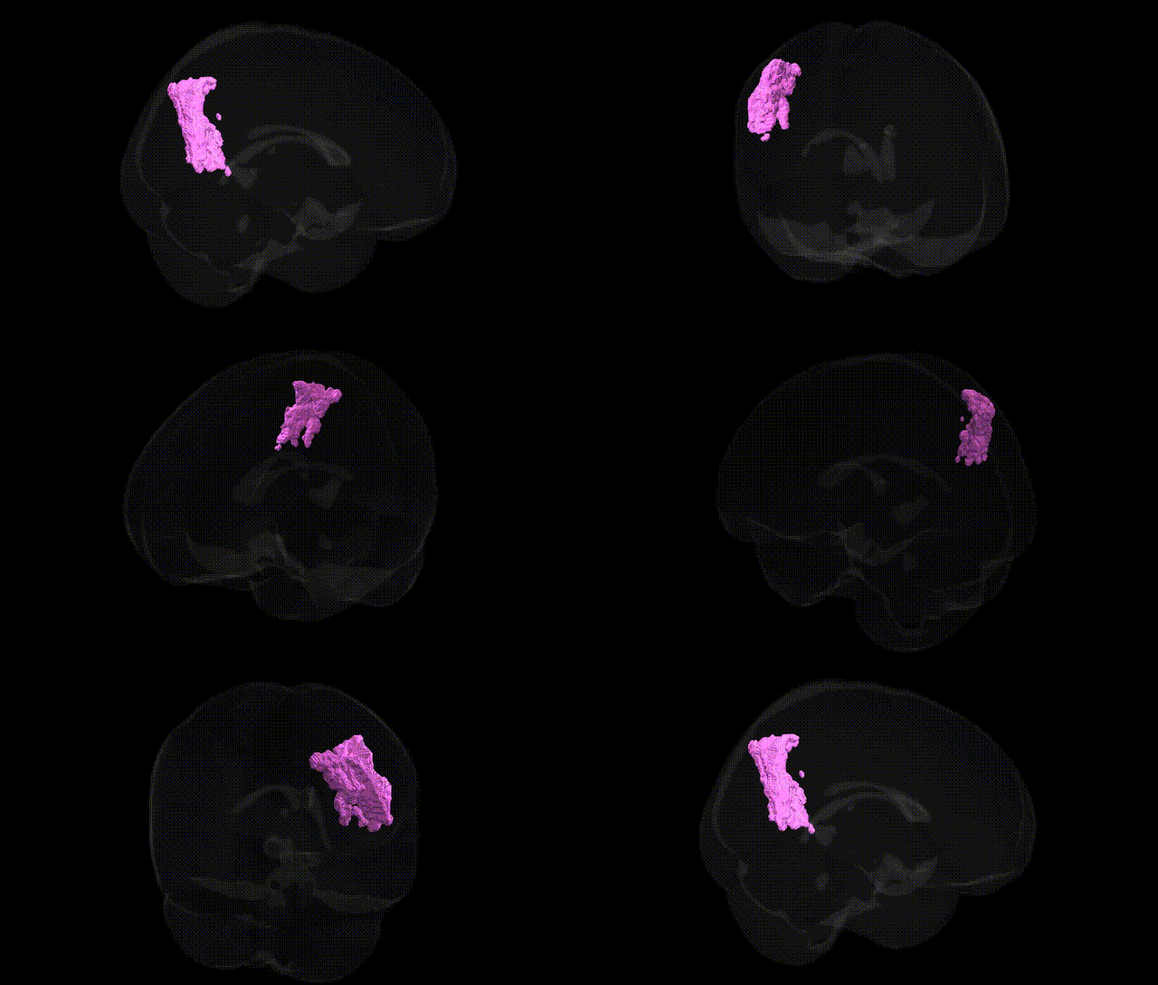
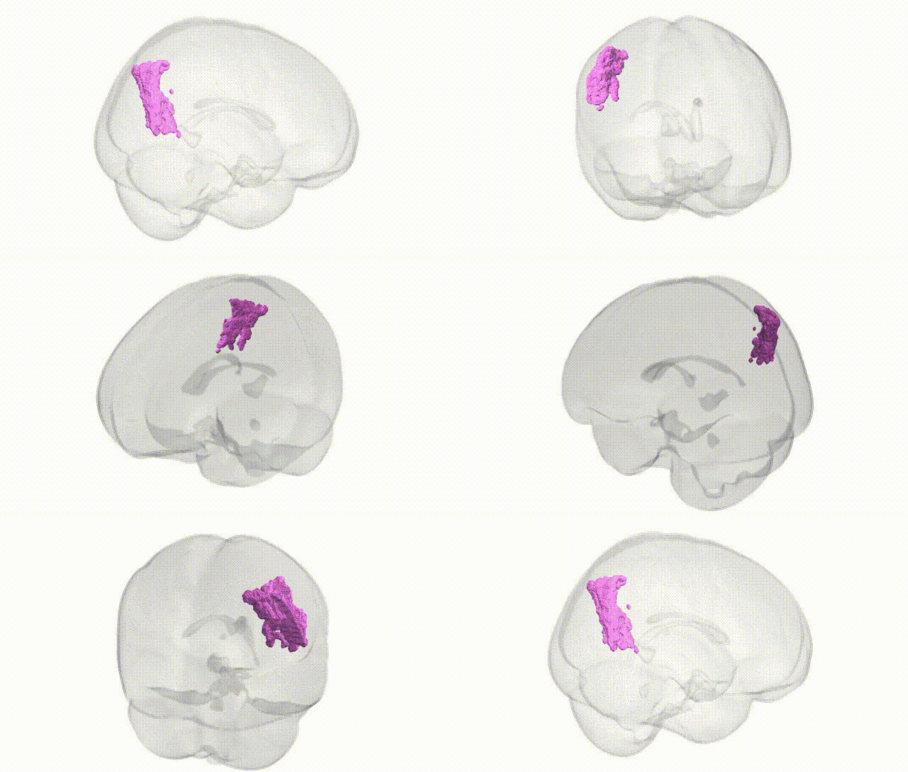
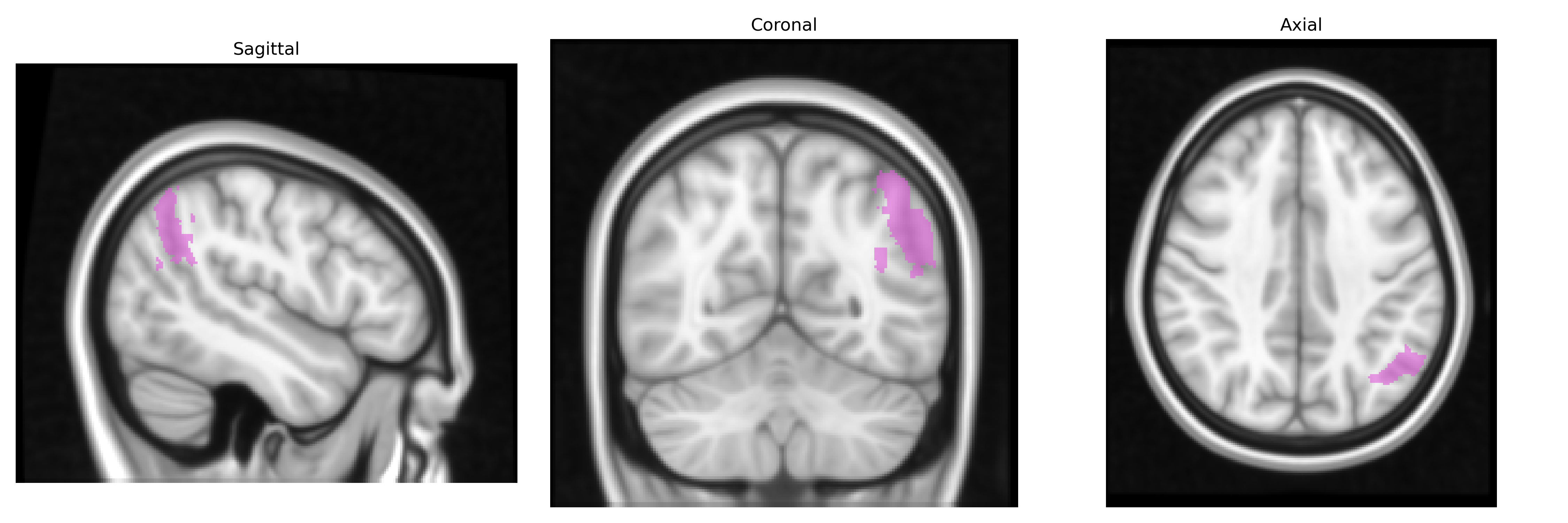
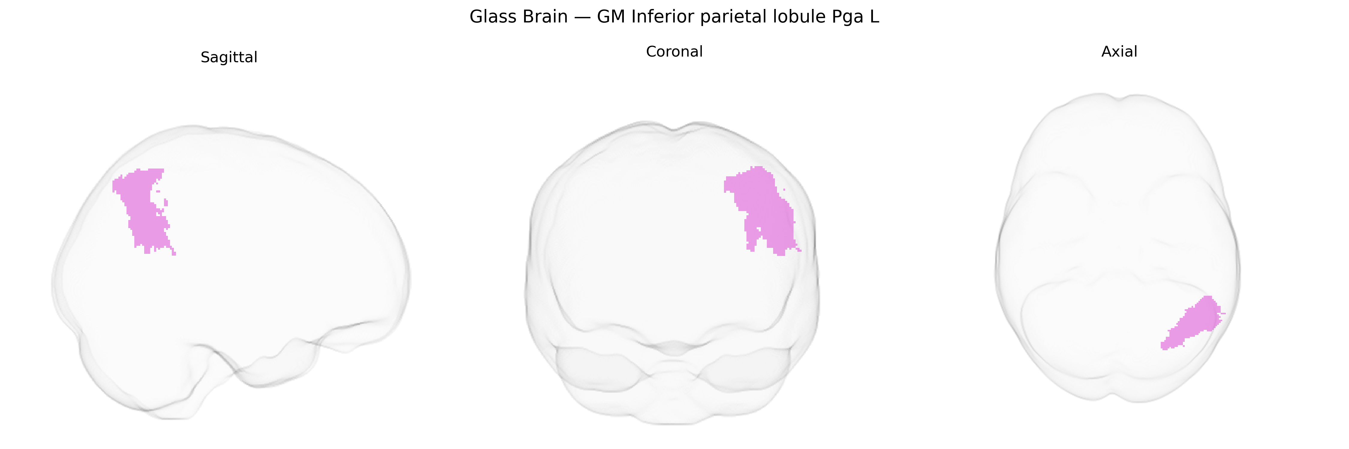
The bilateral GM Inferior parietal lobule PGa L, as defined in the Juelich maxprob thr25 1mm atlas, corresponds to the left-hemispheric granular subdivision PGa of the inferior parietal lobule (IPL), a heteromodal association cortex located in the posterior parietal lobe at the junction of the temporal and occipital lobes. This region is cytoarchitectonically characterized by pronounced granular layers and is implicated in multimodal integration of visual, auditory, and somatosensory information, contributing to higher-order cognitive functions such as language processing (including semantic integration), attention reorientation, working memory, and aspects of social cognition. Functionally, PGa within the IPL participates in large-scale networks including the default mode and frontoparietal control networks, supporting processes like perspective taking, conceptual combination, and the integration of external stimuli with internal representations. There is no direct Wikipedia article for the PGa subregion; a closely related structure is the Inferior parietal lobule.
The bilateral inferior parietal lobule (IPL), including the Juelich-defined GM Inferior parietal lobule Pga L region, has been repeatedly implicated in genetic studies of cognition, language, and psychiatric risk, with many findings derived from large-scale imaging genetics consortia such as ENIGMA and UK Biobank analyses. Polygenic scores for general cognitive ability, educational attainment (heavily influenced by loci near genes such as MAPT, KDM4A, and others across the genome), and language-related traits show robust associations with IPL cortical thickness and surface area, while common variants in and around FOXP2, CNTNAP2, and DCDC2 have been linked to structural and functional alterations in left inferior parietal and perisylvian language networks, including regions overlapping Pga L, in developmental language disorder and dyslexia. Genome-wide association studies of schizophrenia, major depressive disorder, bipolar disorder, and autism spectrum disorder consistently identify polygenic risk effects on IPL structure and connectivity, with implicated loci including calcium channel genes (e.g., CACNA1C), synaptic genes (e.g., GRIN2A, GRM3), and immune-related loci within the MHC region, suggesting that synaptic plasticity and neurodevelopmental pathways shape IPL morphology and function. In Alzheimer’s disease and related dementias, APOE ε4 status and AD polygenic risk scores correlate with atrophy, hypometabolism, and disrupted connectivity in inferior parietal regions that align with Pga L, particularly within the default mode network, while GWAS-identified loci such as BIN1, CLU, PICALM, and SORL1 have been associated with parietal involvement in amyloid and tau-related neurodegeneration. Additional GWAS and candidate gene work link IPL measures to attention-deficit/hyperactivity disorder, reading and mathematical abilities, and visuospatial skills, supporting the view that this region functions as a convergence zone where distributed genetic influences on language, attention, working memory, and neuropsychiatric vulnerability manifest in shared structural and functional variation.
Overview generated by GPT-4o (2026).
Region ID: 37
Hemisphere: bilateral
Atlas: Juelich maxprob thr25 1mm
Full Quality Version: Download MP4
Full Quality Version: Download MP4
Wali Sidiqyar*, Gaurav Rudravaram*, Elyssa M. McMaster, Trent M. Schwartz, Adam M. Saunders, Kurt G. Schilling, Bennett A. Landman "Introducing SPINS: A Shared Public Visualization Library of Neuroanatomical Structures." Medical Imaging with Deep Learning- short paper
This resource is licensed under CC0 1.0 Universal (Public Domain).