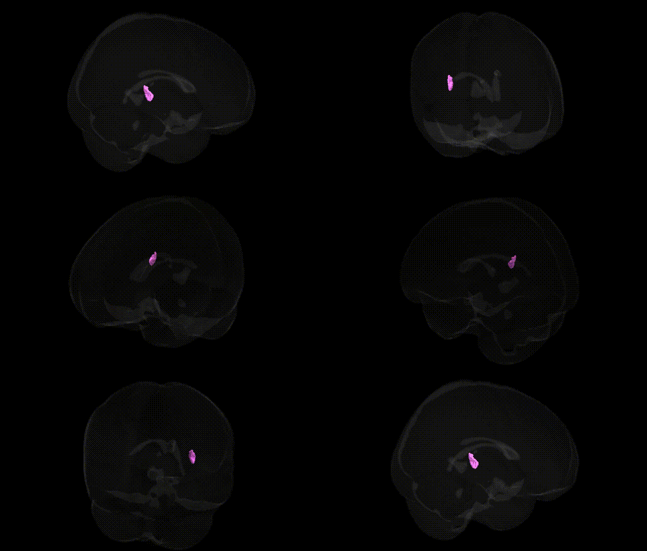
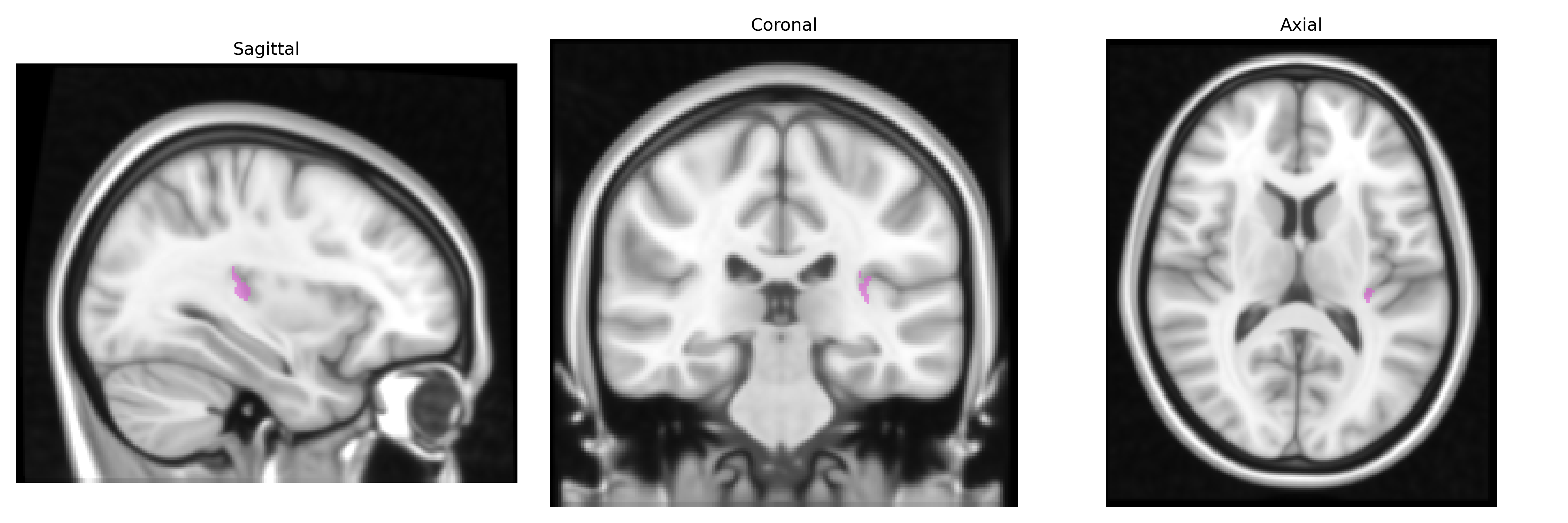
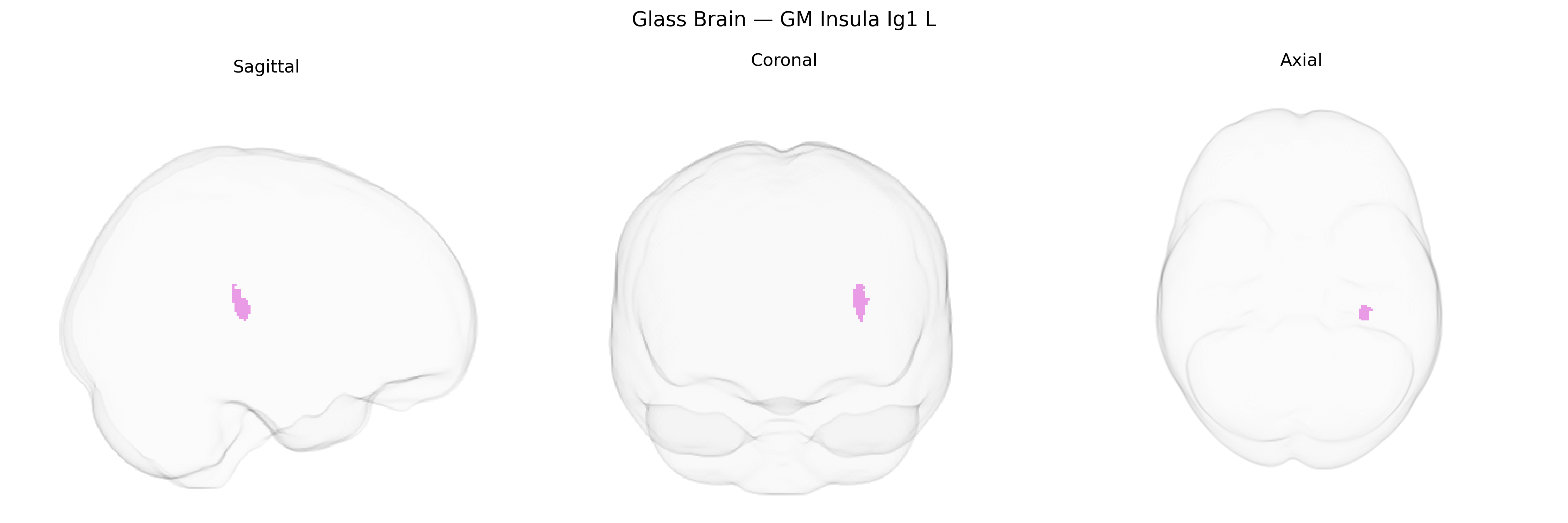
The bilateral GM Insula Ig1 L region in the Juelich maxprob thr25 1 mm atlas corresponds to a granular insular subregion located within the posterior insular cortex, characterized by dense granular layers (particularly layer IV) and strong thalamocortical input. This area is implicated in multimodal sensory integration, particularly somatosensory, visceral, and nociceptive processing, and contributes to interoceptive awareness and autonomic regulation by relaying internal bodily state information to higher-order insular and limbic networks. Functionally, it participates in circuits underlying pain perception, homeostatic control, and aspects of emotional processing, and is interconnected with somatosensory cortices, thalamic nuclei, and other insular fields. There is no direct Wikipedia article for “Insula Ig1”; a related structure is the Insular cortex.
The bilateral GM Insula Ig1 L region in the Juelich maxprob atlas corresponds to anterior/mid-insular cortex, a hub for interoception, salience processing, and affective integration, and has been implicated indirectly in multiple genetic and GWAS-based findings through imaging genetics of insular structure and function. Large-scale GWAS of cortical thickness and surface area (e.g., ENIGMA, UK Biobank) have identified loci in genes such as THBS1, TESC, KIAA0586, and others associated with insular cortical morphology, while polygenic scores for schizophrenia, bipolar disorder, major depression, and autism spectrum disorder correlate with structural and functional alterations in insular subregions, including left anterior insula. Variants in CACNA1C, ZNF804A, and DISC1, commonly implicated in psychosis and mood disorders, show associations with altered insular activation and connectivity during emotional and salience-related tasks. Insular GM measures, including in left insula areas overlapping Ig1, have been used as endophenotypes in GWAS and candidate-gene studies of anxiety, substance use (e.g., nicotine dependence), and pain sensitivity, with genes in stress-response, glutamatergic, and dopaminergic pathways showing effects on insular volume and reactivity. In neurodegenerative and vascular conditions, risk alleles for Alzheimer’s disease (APOE ε4), cerebral small-vessel disease, and stroke have been associated with patterns of atrophy and microstructural change that include insular regions. While no GWAS has yet specifically targeted the Ig1 L parcel itself, convergent imaging-genetic evidence links insular cortex—encompassing Ig1—to polygenic liability for psychiatric disorders, interoceptive and affective traits, and global brain development and aging.
Overview generated by GPT-4o (2026).
Region ID: 118
Hemisphere: bilateral
Atlas: Juelich maxprob thr25 1mm
Full Quality Version: Download MP4
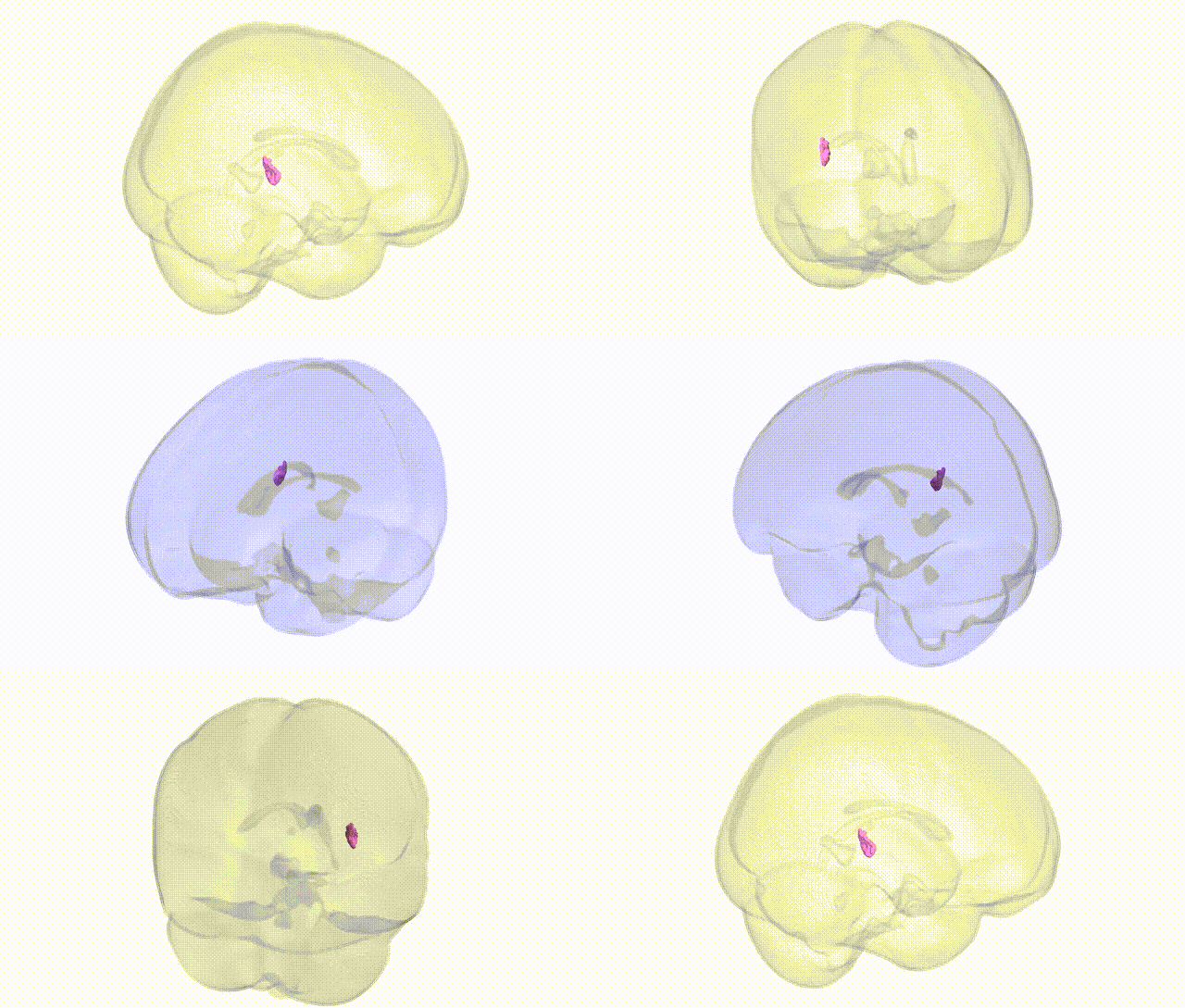
Full Quality Version: Download MP4
Wali Sidiqyar*, Gaurav Rudravaram*, Elyssa M. McMaster, Trent M. Schwartz, Adam M. Saunders, Kurt G. Schilling, Bennett A. Landman "Introducing SPINS: A Shared Public Visualization Library of Neuroanatomical Structures." Medical Imaging with Deep Learning- short paper
This resource is licensed under CC0 1.0 Universal (Public Domain).