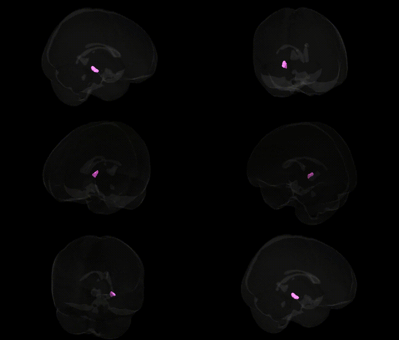
The bilateral GM Lateral geniculate body L in the Juelich maxprob thr25 1mm Atlas represents the left lateral geniculate nucleus (LGN) of the thalamus, a key relay center in the visual pathway where retinal ganglion cell inputs from the contralateral visual field are processed and transmitted to the primary visual cortex (V1). This structure exhibits a characteristic laminar organization, with magnocellular and parvocellular layers that support parallel processing of motion, luminance, and color/form information, and is critical for spatial and temporal aspects of visual perception. The LGN integrates retinal signals with modulatory inputs from cortex and brainstem, contributing to attentional gating and the regulation of visual signal flow. Lateral geniculate nucleus
The lateral geniculate body (LGB), the thalamic relay nucleus for visual input, has been implicated in several genetic and GWAS-based findings, although often via imaging genetics rather than region-specific GWAS. Variants influencing visual system development and integrity, such as in PAX6, SHH, and SOX2, are associated with congenital visual pathway malformations that include the LGN/LGB, while risk alleles for neurodevelopmental disorders (e.g., in CNTNAP2, NRXN1, and synaptic genes) and schizophrenia-associated loci have been linked to structural and functional alterations in thalamic visual nuclei, including the lateral geniculate region. Imaging genetics studies using large cohorts (e.g., UK Biobank) show that common variants associated with overall thalamic volume and white-matter integrity (such as in genes related to axonal guidance, myelination, and synaptic plasticity) can influence the integrity of thalamo-occipital visual pathways that encompass the LGB, and some of these variants overlap with loci implicated in psychiatric disorders, cognitive traits, and sleep regulation. Rare monogenic conditions affecting glutamatergic transmission or GABAergic interneuron development (for example, in GRIN and GAD1-related pathways) have also been reported to alter thalamic sensory nuclei, including the LGN/LGB, contributing to visual processing deficits and broader sensory integration abnormalities. However, as of current knowledge, no large-scale GWAS has identified common variants uniquely and specifically associated with bilateral GM lateral geniculate body volume in the Juelich maxprob thr25 1 mm atlas; genetic associations are inferred mainly from broader thalamic, visual system, and neuropsychiatric imaging genetics findings rather than region-exclusive signals.
Overview generated by GPT-4o (2026).
Region ID: 104
Hemisphere: bilateral
Atlas: Juelich maxprob thr25 1mm
Full Quality Version: Download MP4
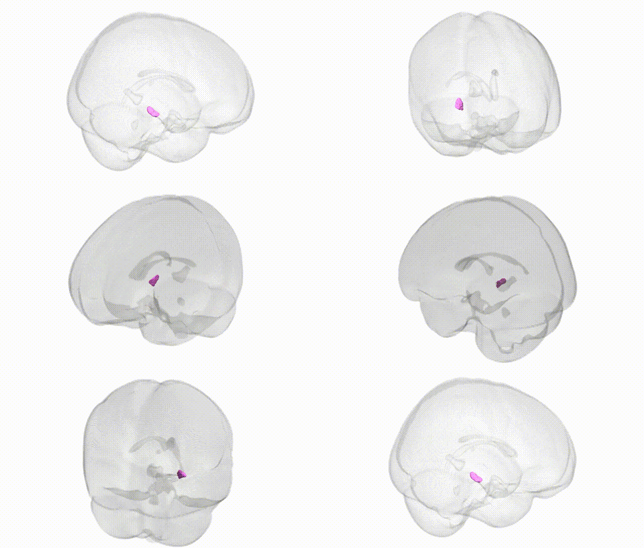
Full Quality Version: Download MP4
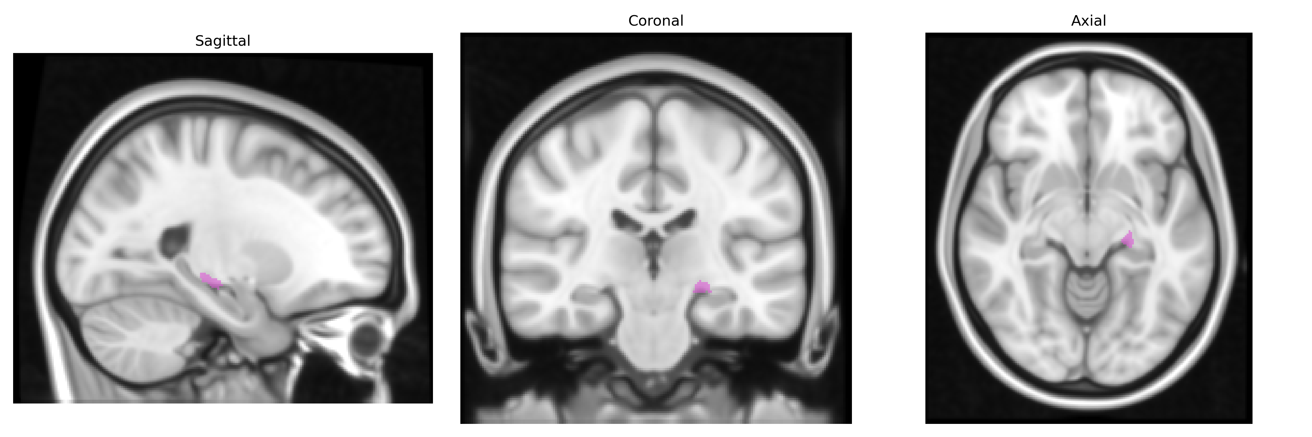
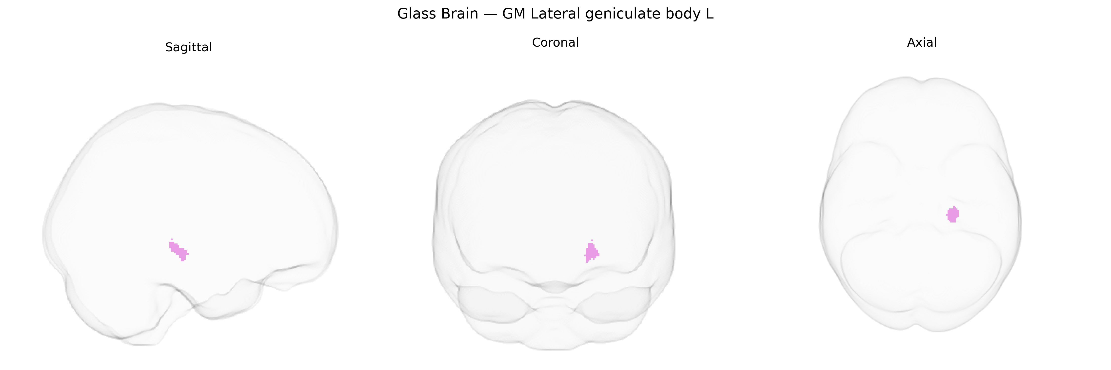
Wali Sidiqyar*, Gaurav Rudravaram*, Elyssa M. McMaster, Trent M. Schwartz, Adam M. Saunders, Kurt G. Schilling, Bennett A. Landman "Introducing SPINS: A Shared Public Visualization Library of Neuroanatomical Structures." Medical Imaging with Deep Learning- short paper
This resource is licensed under CC0 1.0 Universal (Public Domain).