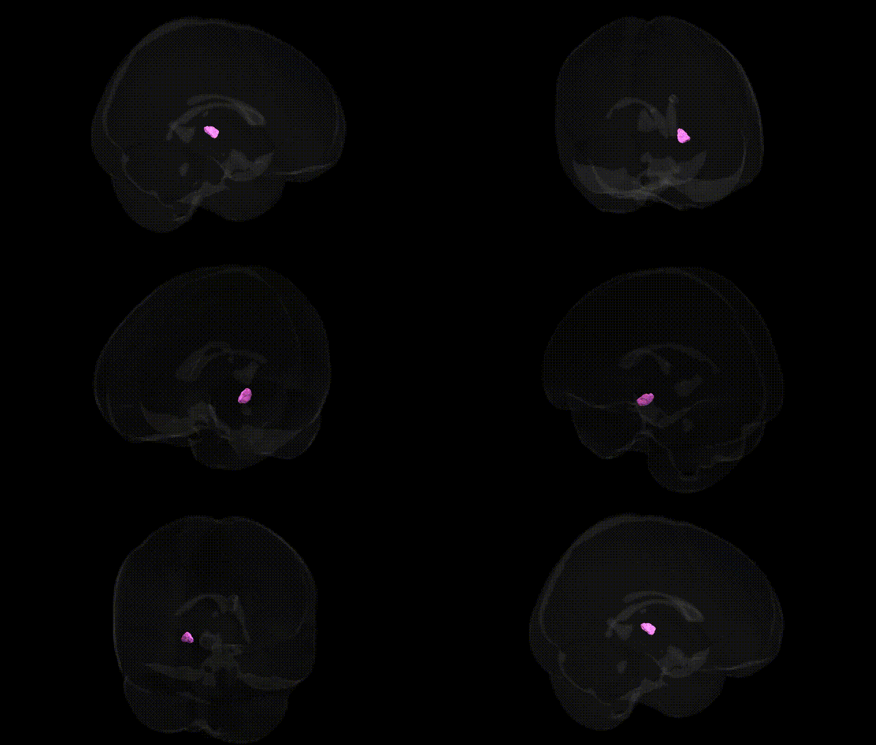
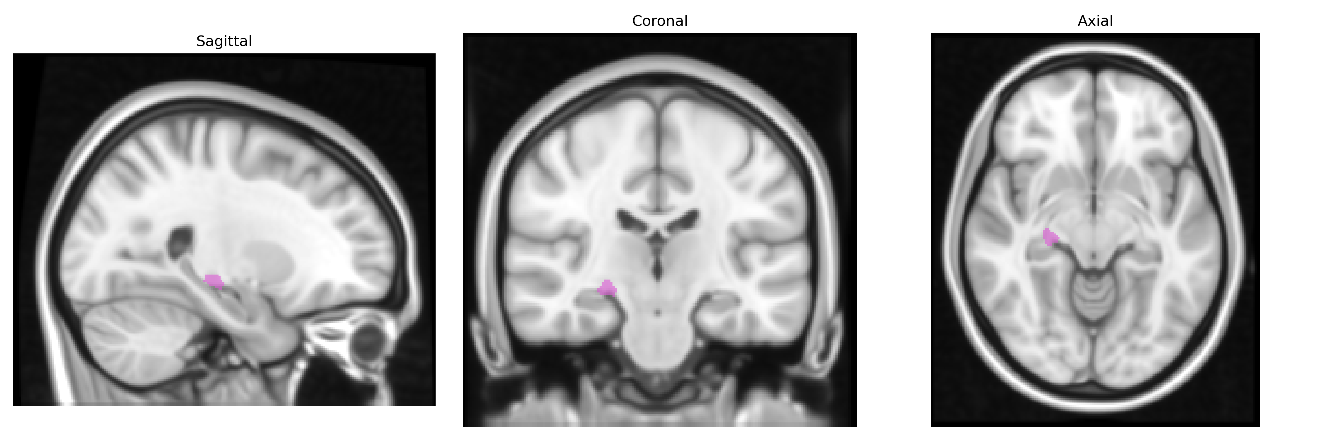
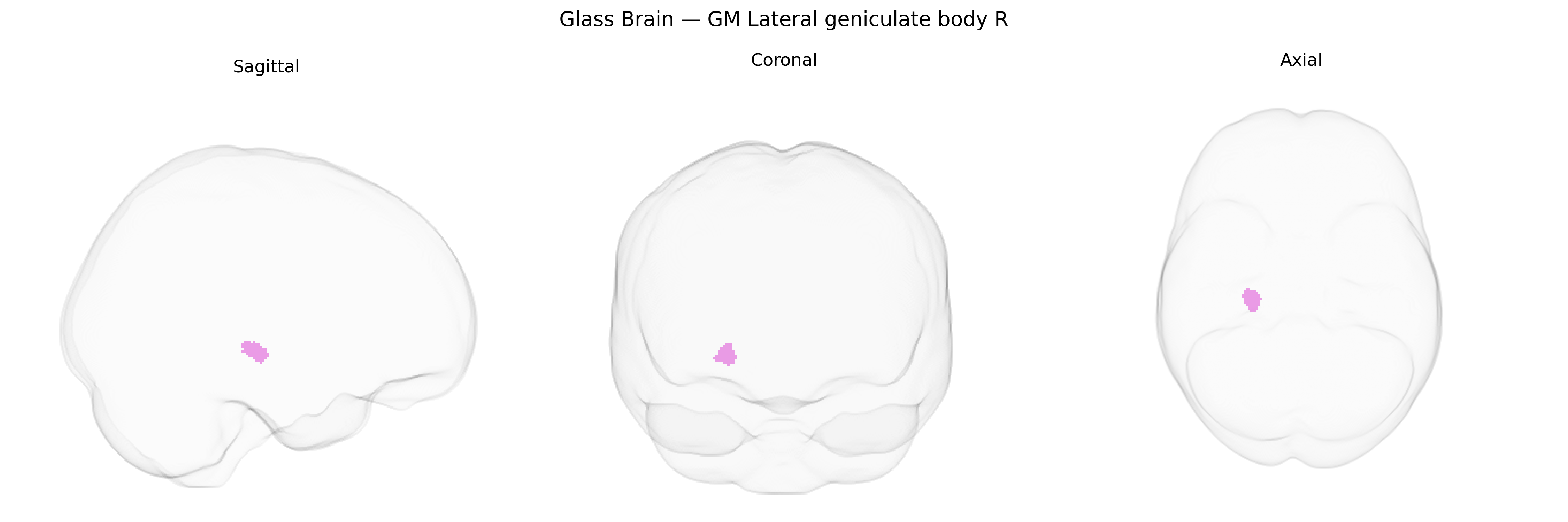
The bilateral GM Lateral geniculate body R, as defined in the Juelich maxprob thr25 1mm atlas, corresponds to the right lateral geniculate nucleus (LGN) of the thalamus, a laminated relay structure in the dorsal thalamus that serves as the primary thalamic relay for visual information from the retina to the primary visual cortex (V1). It receives segregated magnocellular, parvocellular, and koniocellular inputs from retinal ganglion cells via the optic tract and maintains retinotopic organization, enabling parallel processing of motion, luminance contrast, and chromatic/spatial detail. Output from the LGN projects predominantly via the optic radiation to layer IV of the ipsilateral primary visual cortex, preserving eye-specific and spatial mapping. The LGN also receives extensive modulatory feedback from the visual cortex and input from brainstem and other subcortical structures, integrating sensory drive with attentional and state-dependent signals to regulate visual gain and temporal precision of visual transmission. Lateral geniculate nucleus
The lateral geniculate body (LGB), the thalamic relay nucleus for visual input to the cortex, has limited region-specific genetic association data, but several converging findings implicate genes and variants that affect thalamic and visual pathway structure and function. GWAS of subcortical volumes (e.g., ENIGMA) have identified common variants in genes related to neurodevelopment, synaptic function, and axon guidance—such as variants near DCC, PLEKHM1, and genes involved in Wnt and cytoskeletal signaling—that broadly influence thalamic morphology, likely encompassing the LGN/LGB. Polygenic risk scores for schizophrenia, bipolar disorder, and major depression have been associated with thalamic microstructural alterations and thalamo-cortical dysconnectivity, and postmortem and imaging genetics work in psychosis, autism spectrum disorder, and dyslexia repeatedly implicates thalamic visual relay nuclei, including the LGB, in disrupted sensory processing and attentional gating. Variants in genes regulating myelination (e.g., NRG1–ERBB signaling), glutamatergic transmission (e.g., GRIN2A/B), and inhibitory circuitry (e.g., GAD1) have been linked to disorders showing structural or functional LGB abnormalities, such as schizophrenia, bipolar disorder, and visual hallucinations. Additionally, GWAS of visual acuity, refractive error, and retinal morphology (e.g., variants near GJD2, RASGRF1, and other neurodevelopmental genes) implicate pathways that influence the integrity of the retino-geniculo-cortical system, suggesting indirect genetic influences on LGB structure and function, even though no large GWAS to date has reported locus-specific associations uniquely constrained to the Juelich bilateral GM Lateral geniculate body R region.
Overview generated by GPT-4o (2026).
Region ID: 103
Hemisphere: bilateral
Atlas: Juelich maxprob thr25 1mm
Full Quality Version: Download MP4
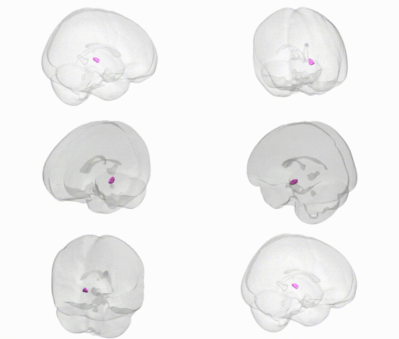
Full Quality Version: Download MP4
Wali Sidiqyar*, Gaurav Rudravaram*, Elyssa M. McMaster, Trent M. Schwartz, Adam M. Saunders, Kurt G. Schilling, Bennett A. Landman "Introducing SPINS: A Shared Public Visualization Library of Neuroanatomical Structures." Medical Imaging with Deep Learning- short paper
This resource is licensed under CC0 1.0 Universal (Public Domain).