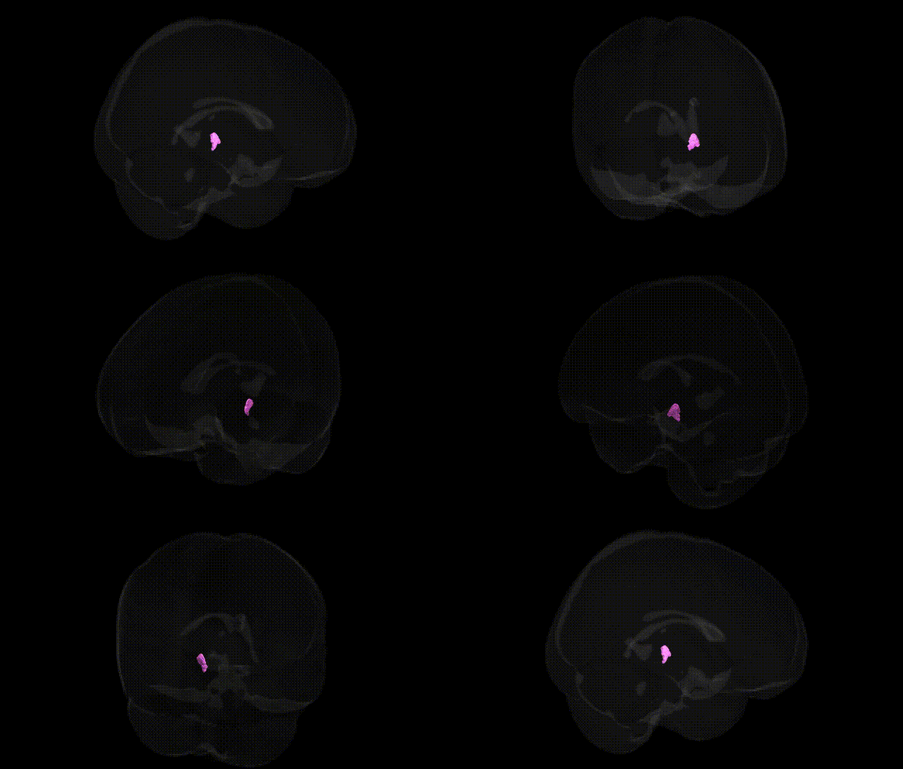
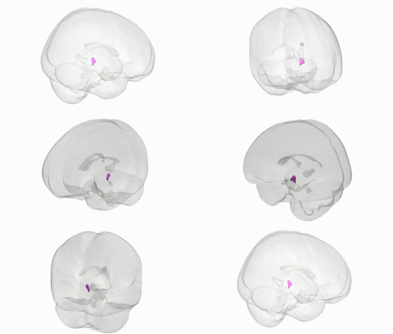
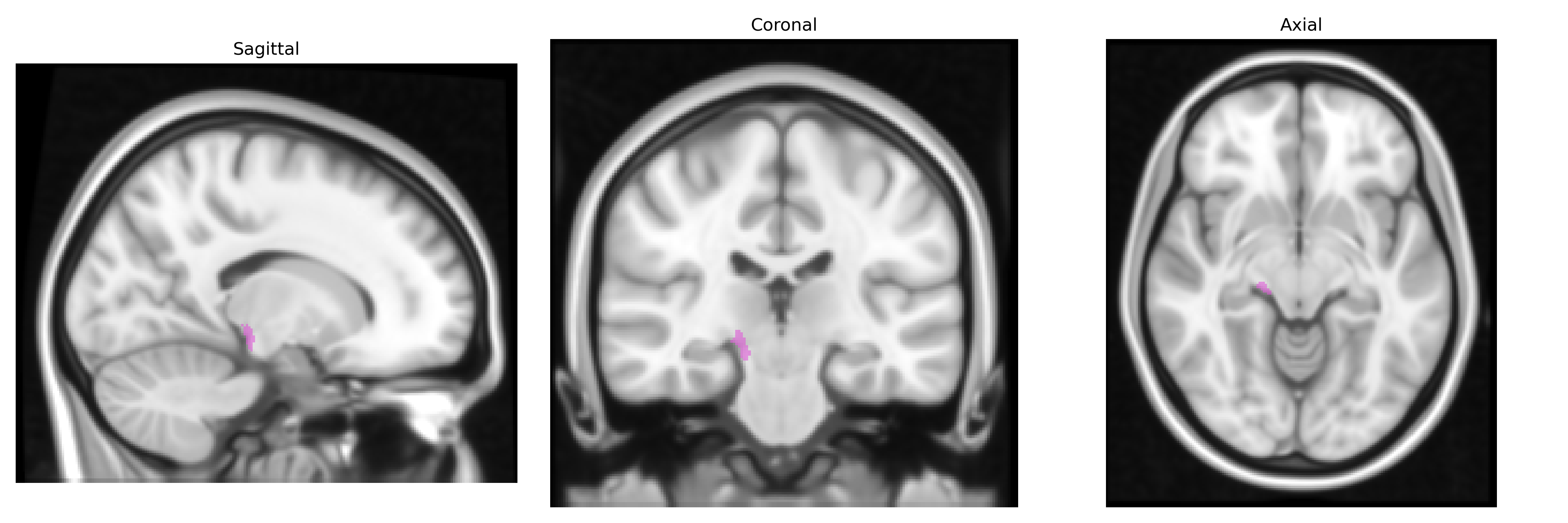
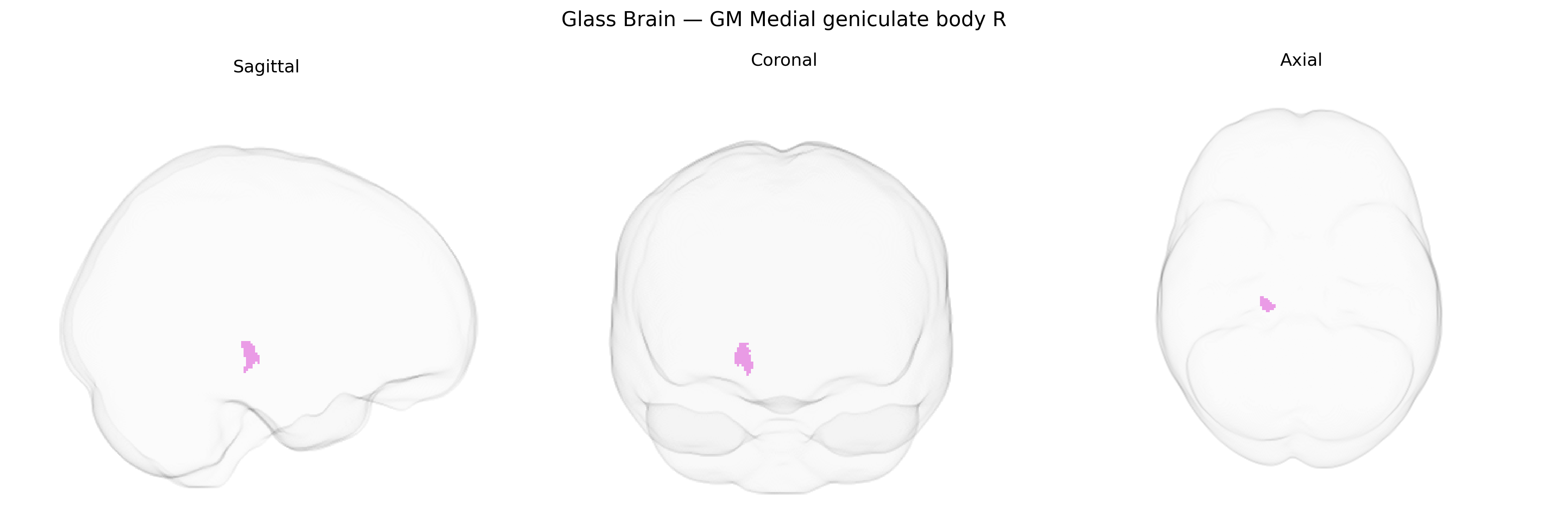
The bilateral GM Medial geniculate body R from the Juelich maxprob thr25 1mm Atlas refers to gray matter of the right medial geniculate nucleus (body), a key thalamic relay center in the auditory pathway. Located in the medial portion of the metathalamus, adjacent to the inferior colliculus and continuous with other thalamic nuclei, it receives ascending auditory input primarily from the inferior colliculus via the brachium of the inferior colliculus and projects to the primary auditory cortex in the temporal lobe. Functionally, this structure participates in frequency, intensity, and temporal processing of sound, contributes to sound localization, and plays a role in selective auditory attention and gating. It is cytoarchitectonically subdivided into ventral, dorsal, and medial divisions with distinct connectivity and response properties, supporting both precise auditory discrimination and integrative multisensory and cognitive aspects of hearing. Medial geniculate nucleus
The medial geniculate body (MGB), the thalamic relay nucleus of the auditory pathway, has no extensively characterized, region-specific GWAS findings from large-scale neuroimaging genetics consortia (e.g., ENIGMA, UK Biobank) that isolate the Juelich maxprob thr25 1 mm bilateral GM Medial geniculate body R region, but genetic studies implicate auditory thalamic structures—including the MGB—in several disorders and traits. Variants affecting glutamatergic and GABAergic signaling (such as in GRM3, GAD1, and related synaptic genes) and thalamocortical circuitry have been associated with schizophrenia, bipolar disorder, and major depression, where abnormal thalamic sensory gating and auditory processing are frequently reported. In tinnitus, candidate-gene and imaging genetics work highlights maladaptive plasticity and hyperexcitability in auditory thalamus, with indirect links to genes involved in neuroplasticity and excitatory/inhibitory balance (e.g., BDNF, GRIN2B), although associations are typically at the system level rather than MGB-specific. Autism spectrum disorder and dyslexia studies have reported altered microstructure and functional connectivity in auditory thalamus, with genetic convergence on synaptic, axon guidance, and neurodevelopmental pathways (e.g., CNTNAP2, FOXP2, DCDC2), again implicating auditory thalamic hubs without fine-grained geniculate localization. Large GWAS of subcortical volumes identify polygenic influences (including loci near HMGA2, FAT3, and others) on thalamic size and shape, and by extension likely affect MGB morphology and function, but to date no robust, replicated SNPs or genes are uniquely tied to the MGB region defined in the Juelich atlas; current genetic links therefore remain indirect, via broader thalamic or auditory network associations.
Overview generated by GPT-4o (2026).
Region ID: 106
Hemisphere: bilateral
Atlas: Juelich maxprob thr25 1mm
Full Quality Version: Download MP4
Full Quality Version: Download MP4
Wali Sidiqyar*, Gaurav Rudravaram*, Elyssa M. McMaster, Trent M. Schwartz, Adam M. Saunders, Kurt G. Schilling, Bennett A. Landman "Introducing SPINS: A Shared Public Visualization Library of Neuroanatomical Structures." Medical Imaging with Deep Learning- short paper
This resource is licensed under CC0 1.0 Universal (Public Domain).