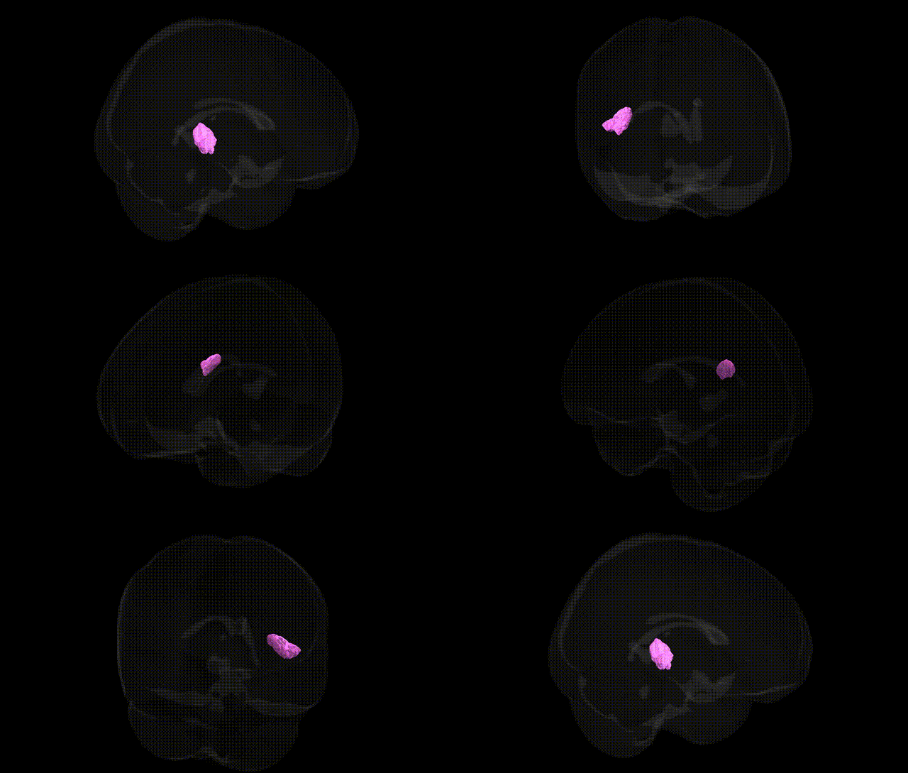
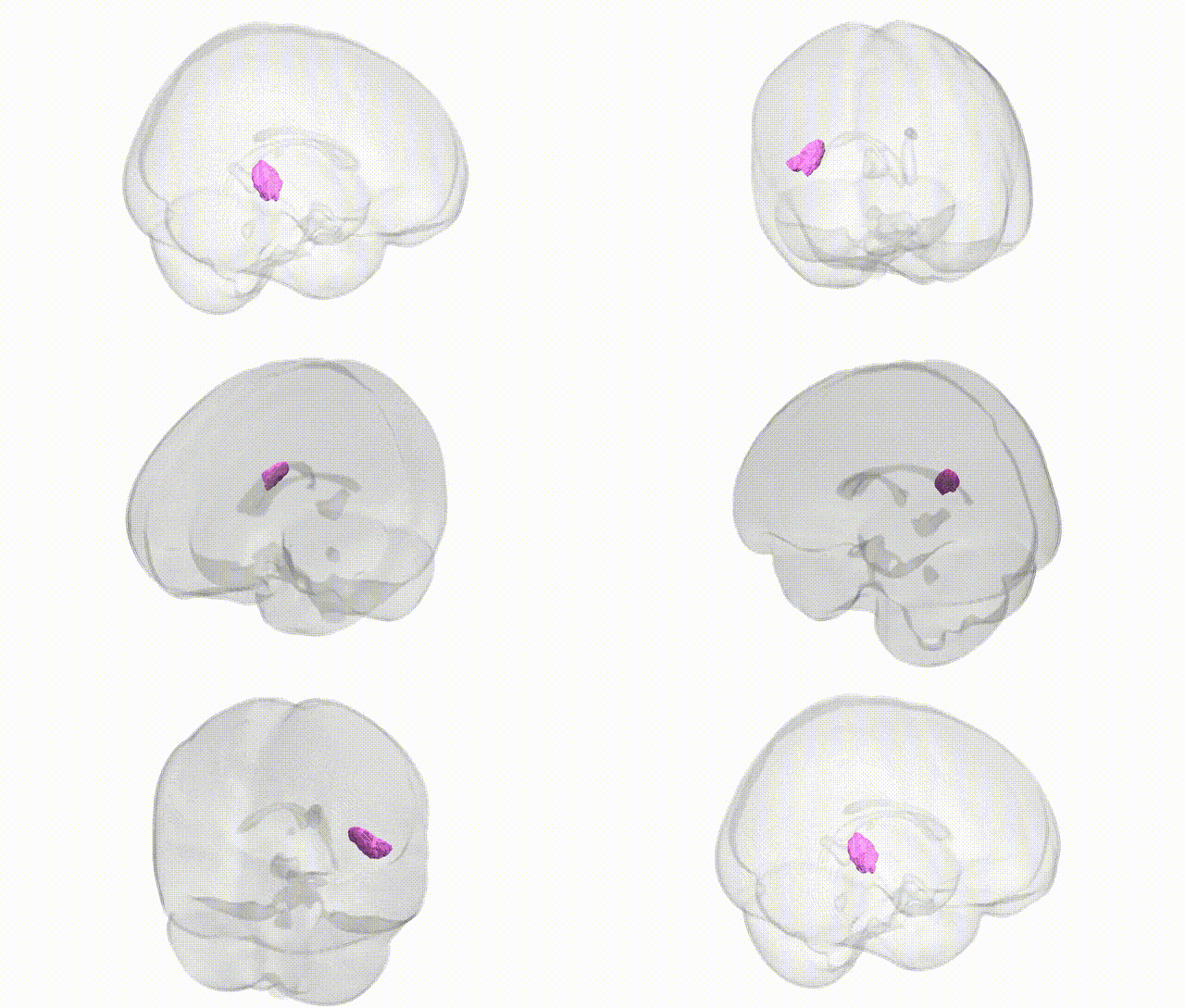
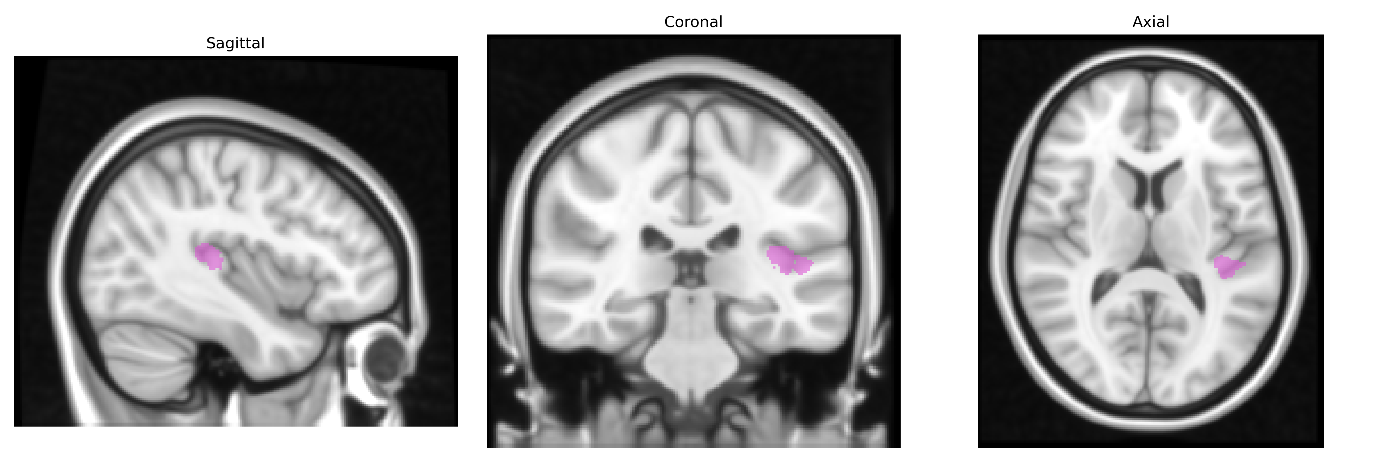
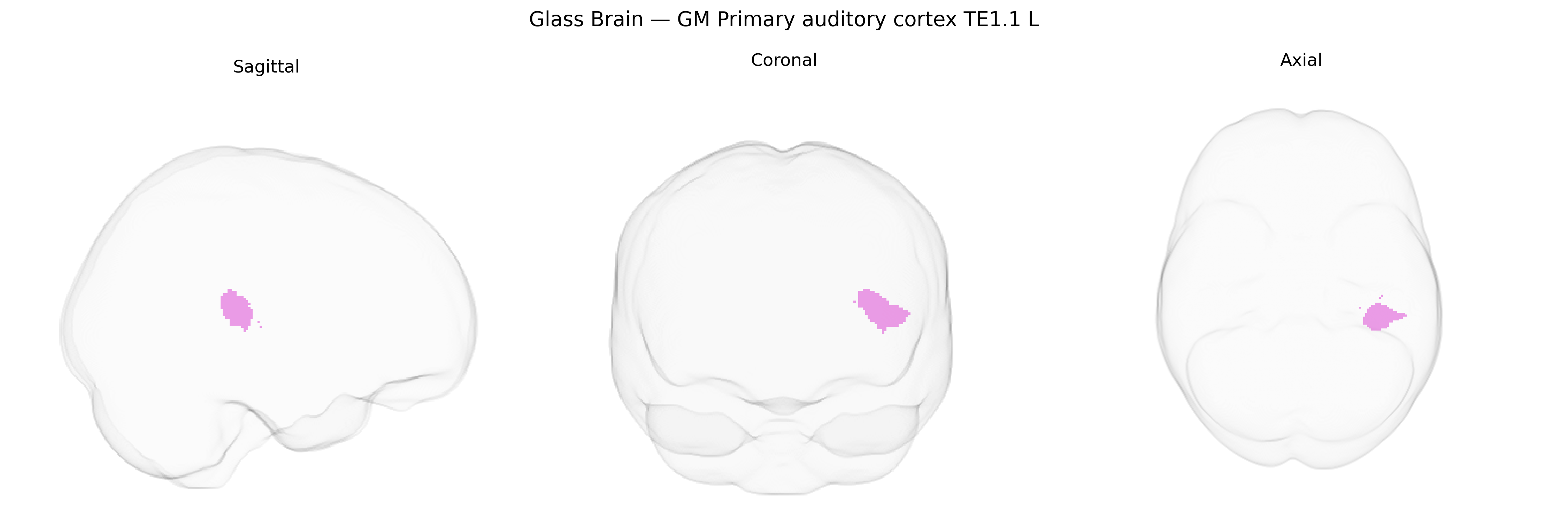
The bilateral GM Primary auditory cortex TE1.1 L, as defined in the Juelich maxprob thr25 1mm atlas, corresponds to a cytoarchitectonically delineated subregion of Heschl’s gyrus forming part of the core primary auditory cortex. It is characterized by a highly granular layer IV, dense thalamocortical (medial geniculate nucleus) input, and tonotopic organization, supporting precise encoding of basic acoustic features such as frequency, intensity, and temporal structure of sounds. This region participates in early-stage auditory processing that underlies more complex functions such as speech and music perception, and it exhibits strong connectivity with adjacent auditory belt and parabelt areas as well as multimodal association cortices. No direct Wikipedia article exists for TE1.1; a related structure is Heschl’s gyrus.
The bilateral grey matter primary auditory cortex TE1.1 L (Juelich maxprob thr25 1mm atlas) corresponds to core auditory cortex within Heschl’s gyrus, a region whose structure and function show heritable variation and multiple genetic associations. Twin and family studies indicate substantial heritability of Heschl’s gyrus volume and cortical thickness, and GWAS of brain imaging phenotypes (e.g., ENIGMA, UK Biobank) have linked variants in genes involved in synaptic function, neuronal migration, and axon guidance (such as those in glutamatergic and GABAergic pathways) to microstructural measures in primary auditory cortex, although individual locus-level findings are still emerging and often subthreshold. Genetic studies of reading and language disorders (including developmental dyslexia and specific language impairment) have implicated genes such as DCDC2, KIAA0319, and ROBO1, which are associated with atypical morphology or activation patterns in left Heschl’s gyrus and adjacent auditory areas during phonological processing. GWAS of tinnitus and hearing-related traits have identified loci (e.g., in genes like PTPRD, GRM7, and others involved in auditory transduction and central processing) that show functional and connectivity links to primary auditory cortex, consistent with central gain and maladaptive plasticity models of tinnitus. Additionally, schizophrenia, autism spectrum conditions, and hallucination-prone phenotypes exhibit genetically influenced alterations in primary auditory cortex structure and auditory evoked responses, with risk variants in neurodevelopmental and synaptic genes (including CACNA1C, GRIN2A, and CNTNAP2 among others) associated with differences in temporal cortex and auditory network organization that encompass TE1.1.
Overview generated by GPT-4o (2026).
Region ID: 43
Hemisphere: bilateral
Atlas: Juelich maxprob thr25 1mm
Full Quality Version: Download MP4
Full Quality Version: Download MP4
Wali Sidiqyar*, Gaurav Rudravaram*, Elyssa M. McMaster, Trent M. Schwartz, Adam M. Saunders, Kurt G. Schilling, Bennett A. Landman "Introducing SPINS: A Shared Public Visualization Library of Neuroanatomical Structures." Medical Imaging with Deep Learning- short paper
This resource is licensed under CC0 1.0 Universal (Public Domain).