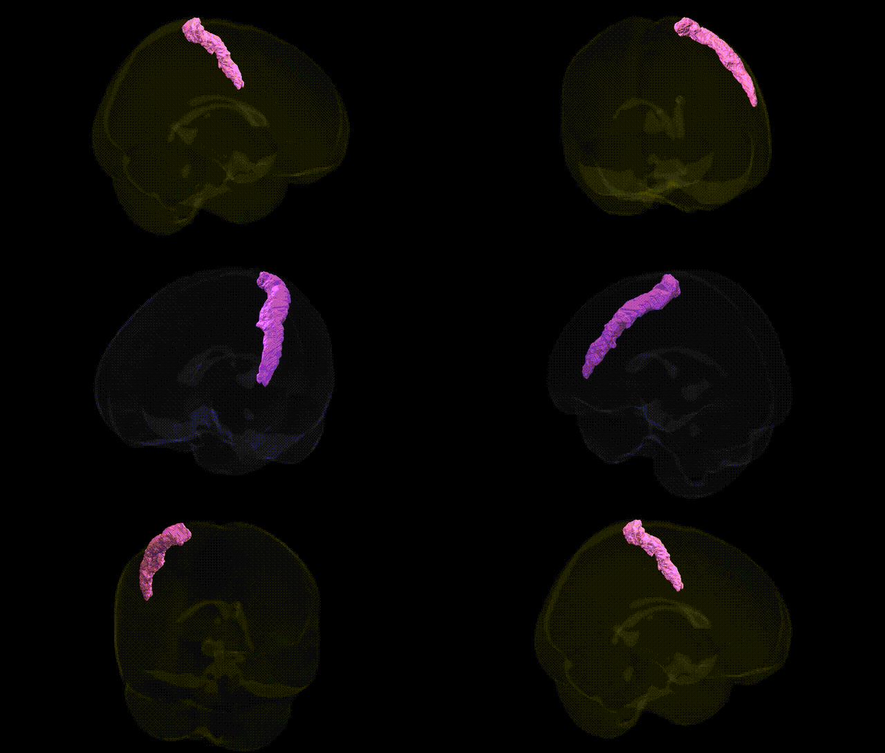
The bilateral GM Primary somatosensory cortex BA1 R, as defined in the Juelich maxprob thr25 1mm Atlas, corresponds to Brodmann area 1 within the postcentral gyrus of the parietal lobe, predominantly located on the crown of the gyrus posterior to the central sulcus. This region forms part of the primary somatosensory cortex (SI) and is specialized for processing cutaneous sensory input, including fine touch, texture, and aspects of vibratory and rapidly adapting mechanoreceptive signals. BA1 receives dense thalamocortical input from the ventral posterior nucleus and integrates information relayed from adjacent somatosensory fields (notably BA3b and BA2), contributing to the construction of somatotopically organized representations of the body surface. Neuronal populations in BA1 exhibit columnar organization and modality-specific response properties, supporting higher-order discrimination and perception of tactile stimuli necessary for sensorimotor integration and object manipulation. No direct Wikipedia article exists for “Brodmann area 1” as a standalone entry; a closely related and encompassing structure is the Primary somatosensory cortex.
The bilateral gray matter primary somatosensory cortex BA1, as defined in the Juelich maxprob thr25 1mm atlas, has been implicated indirectly in genetic studies linking common variants to sensorimotor processing, pain perception, and neurodevelopmental traits, though few GWAS report locus-specific associations explicitly to BA1. Large-scale brain imaging GWAS (e.g., ENIGMA, UK Biobank) have identified variants in genes such as HMGA2, IGF1, CENPW, and those in neurodevelopmental and synaptic pathways (e.g., MAPT region, glutamatergic signaling genes) that modulate cortical thickness and surface area in somatosensory and adjacent sensorimotor cortices, suggesting shared polygenic influences on BA1 structure. Somatosensory cortex, including BA1, shows altered morphology and activation in chronic pain conditions (e.g., migraine, neuropathic pain, fibromyalgia), for which GWAS have highlighted genes involved in nociception and neuroinflammation (such as CACNA1A, TRPM8, and HLA-region variants), implying that genetic variation in pain pathways may exert downstream effects on BA1 function. Neurodevelopmental and psychiatric disorders with strong genetic components—autism spectrum disorder, ADHD, schizophrenia—frequently show atypical somatosensory cortex organization or connectivity, with GWAS pointing to synaptic, chromatin-remodeling, and neuronal migration genes (e.g., CACNA1C, GRIA1, FOXP1, CHD8) that broadly impact cortical circuit formation, likely including BA1. Additionally, twin and family studies indicate high heritability for somatosensory-evoked potentials and tactile acuity, consistent with a polygenic architecture influencing BA1 excitability and microstructure, even if region-specific genetic effects are currently mapped at relatively coarse spatial resolution and not uniquely resolved to BA1 in most GWAS catalogs.
Overview generated by GPT-4o (2026).
Region ID: 52
Hemisphere: bilateral
Atlas: Juelich maxprob thr25 1mm
Full Quality Version: Download MP4
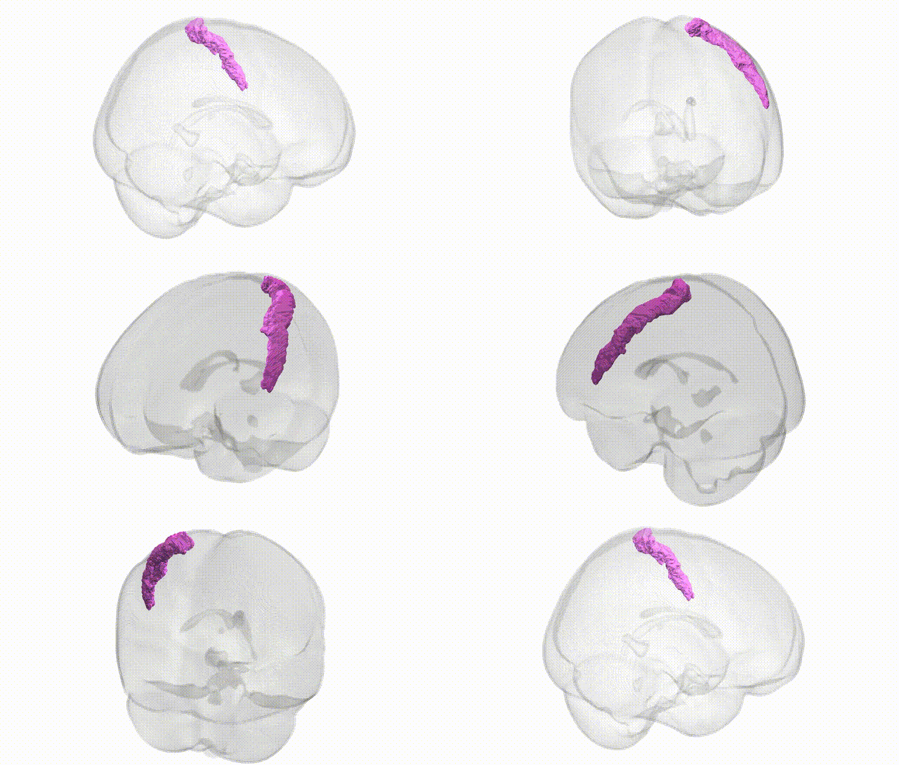
Full Quality Version: Download MP4
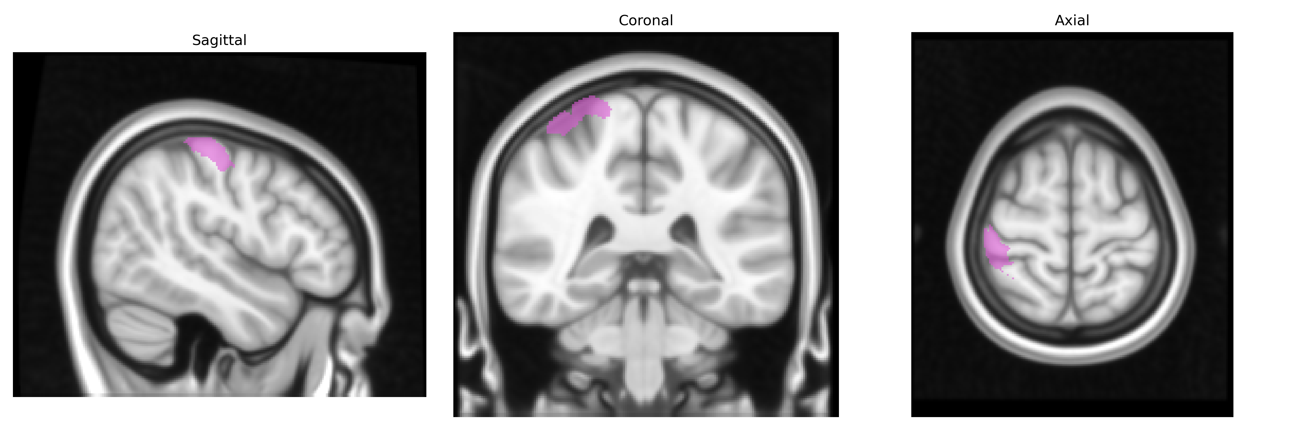
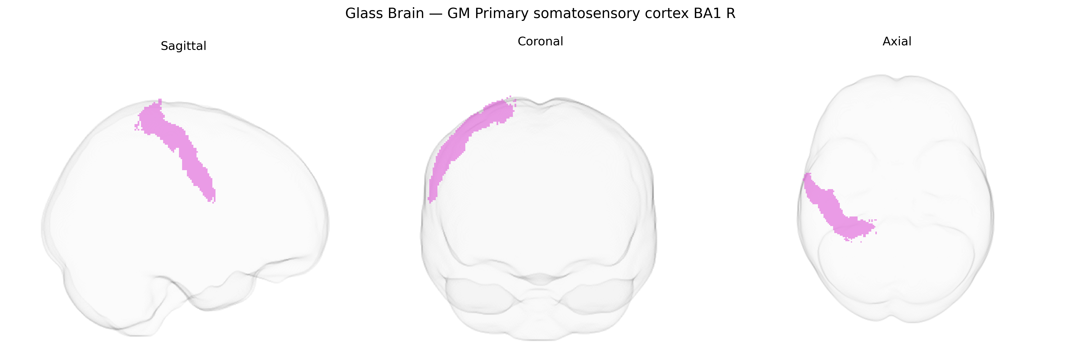
Wali Sidiqyar*, Gaurav Rudravaram*, Elyssa M. McMaster, Trent M. Schwartz, Adam M. Saunders, Kurt G. Schilling, Bennett A. Landman "Introducing SPINS: A Shared Public Visualization Library of Neuroanatomical Structures." Medical Imaging with Deep Learning- short paper
This resource is licensed under CC0 1.0 Universal (Public Domain).