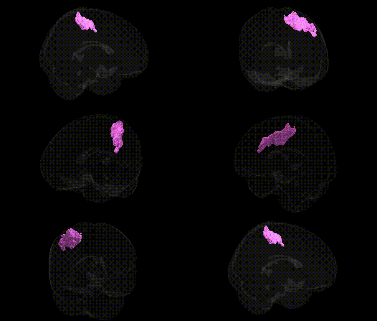
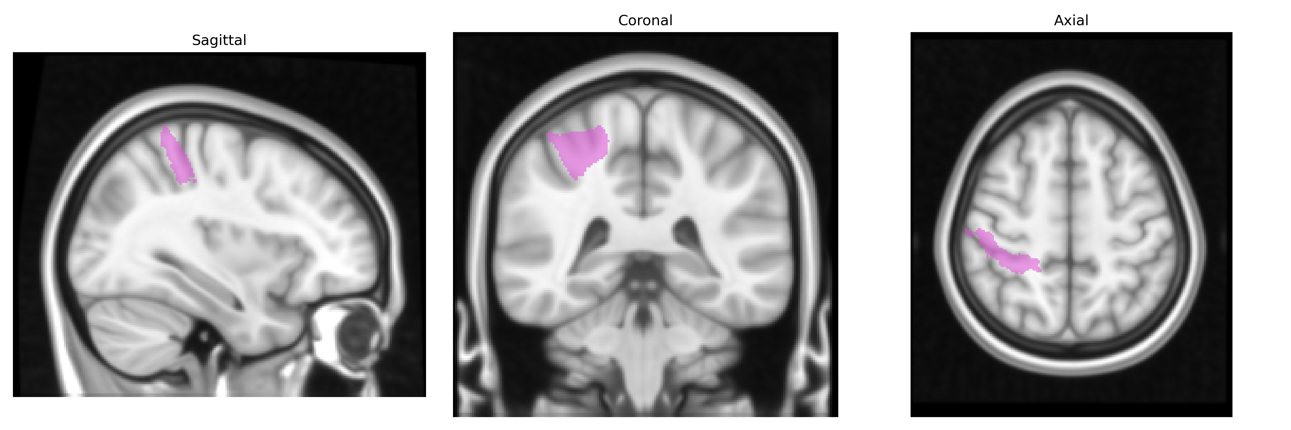
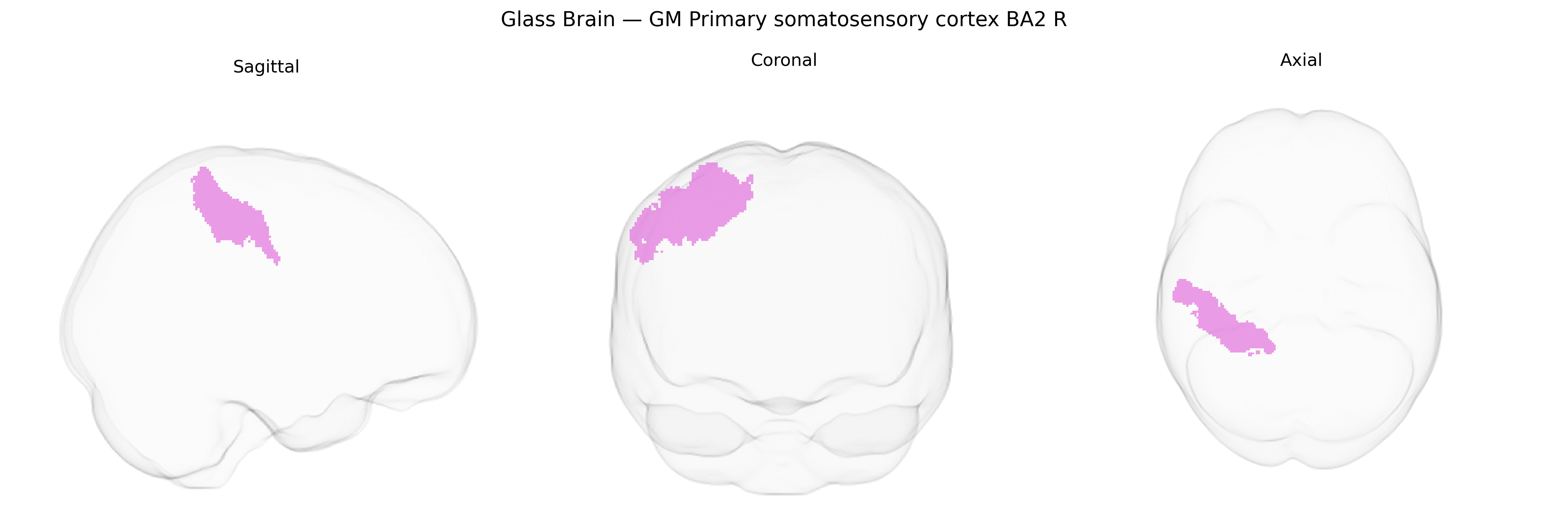
The bilateral GM Primary somatosensory cortex BA2 R, as defined in the Juelich maxprob thr25 1mm Atlas, corresponds to the granular field Brodmann area 2 located in the postcentral gyrus of the parietal lobe, primarily on the right hemisphere but represented bilaterally in this label. BA2 is part of the primary somatosensory cortex (S1) and receives dense input from thalamic relay nuclei via earlier somatosensory areas (BA3 and BA1), integrating tactile, proprioceptive, and kinesthetic information to support higher-order processing of object shape, size, and limb position. Neuronal populations in BA2 exhibit complex receptive fields and contribute to sensorimotor integration, fine discrimination of tactile stimuli, and coordinated motor planning through reciprocal connections with secondary somatosensory and motor areas. Related article: Brodmann area 2.
Genetic associations specific to the bilateral GM Primary somatosensory cortex BA2 R (as defined in the Juelich maxprob thr25 1mm atlas) are not extensively characterized at the fine-grained regional level, but broader GWAS and imaging-genetics studies implicating the primary somatosensory cortex and adjacent parietal regions provide relevant context. Large-scale brain MRI GWAS (e.g., ENIGMA, UK Biobank) have identified variants in loci such as HMGA2, MIR137, and multiple glutamatergic and neurodevelopmental genes that influence cortical thickness, surface area, and gyrification in somatosensory and parietal cortices, suggesting polygenic control of BA2 structure. Somatosensory cortex involvement is reported in genetic studies of chronic pain (e.g., migraine, back pain, neuropathic pain), in which risk variants in genes related to neuronal excitability (CACNA1A, SCN9A), inflammatory signaling, and synaptic plasticity are associated with altered somatosensory processing and functional activation. Neurodevelopmental and neuropsychiatric GWAS, including those for autism spectrum disorder, ADHD, and schizophrenia, have identified risk loci in synaptic and cortical development genes (such as CNTNAP2, NRXN1, and GRIN2A) that correlate with atypical somatosensory cortex structure or function in imaging-genetics follow-up analyses, although these associations are typically reported at the lobar (parietal) or network level rather than BA2 specifically. Additionally, variants linked to handedness and motor skill (e.g., in PCSK6 and other lateralization-related genes) have shown associations with sensorimotor cortical asymmetries, indirectly implicating BA2. Overall, genetic influences on BA2 R are inferred from broader somatosensory and parietal cortex GWAS, chronic pain genetics, and neurodevelopmental disorder imaging-genetics, but precise BA2 R–specific loci remain incompletely resolved in current literature.
Overview generated by GPT-4o (2026).
Region ID: 54
Hemisphere: bilateral
Atlas: Juelich maxprob thr25 1mm
Full Quality Version: Download MP4
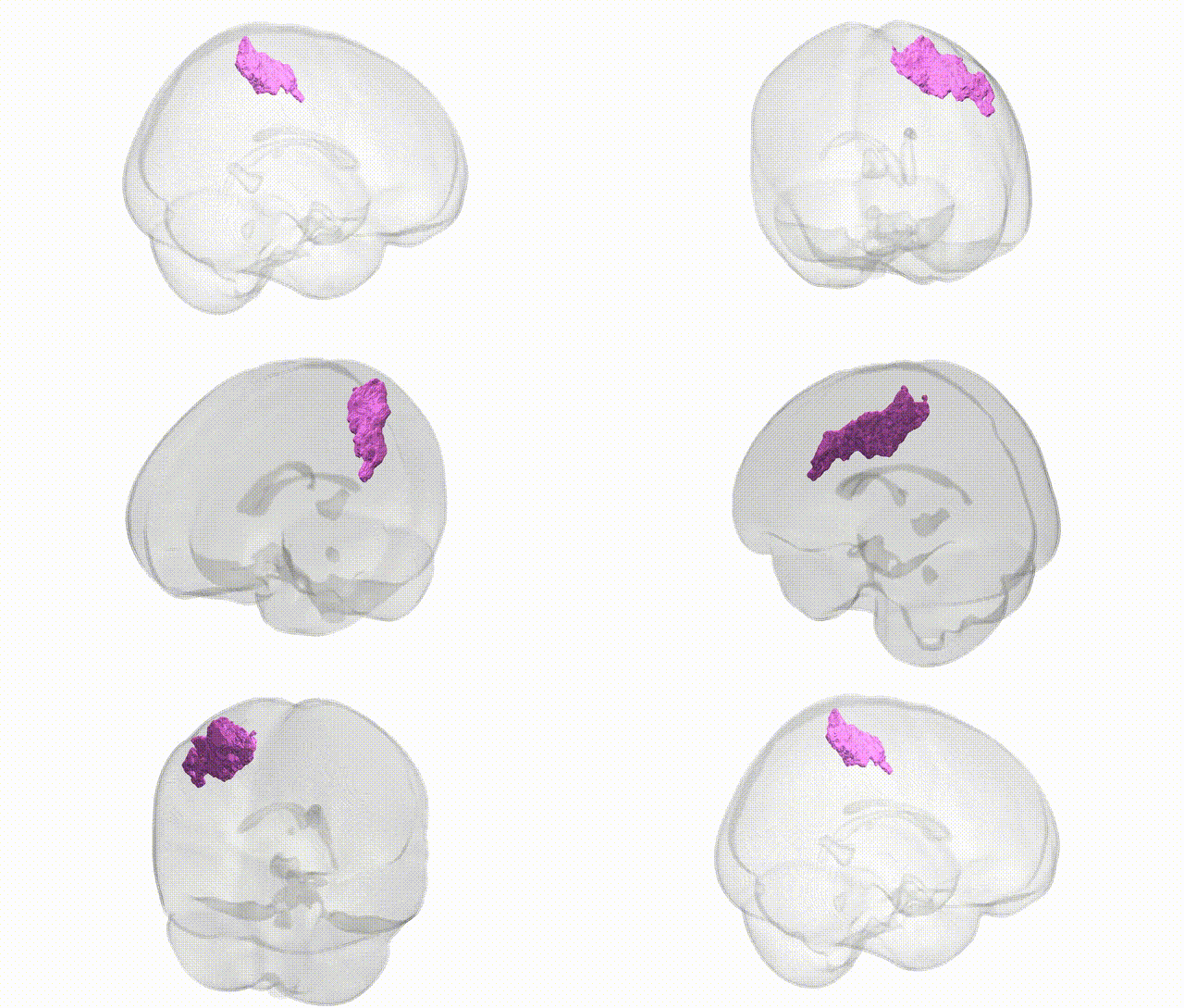
Full Quality Version: Download MP4
Wali Sidiqyar*, Gaurav Rudravaram*, Elyssa M. McMaster, Trent M. Schwartz, Adam M. Saunders, Kurt G. Schilling, Bennett A. Landman "Introducing SPINS: A Shared Public Visualization Library of Neuroanatomical Structures." Medical Imaging with Deep Learning- short paper
This resource is licensed under CC0 1.0 Universal (Public Domain).