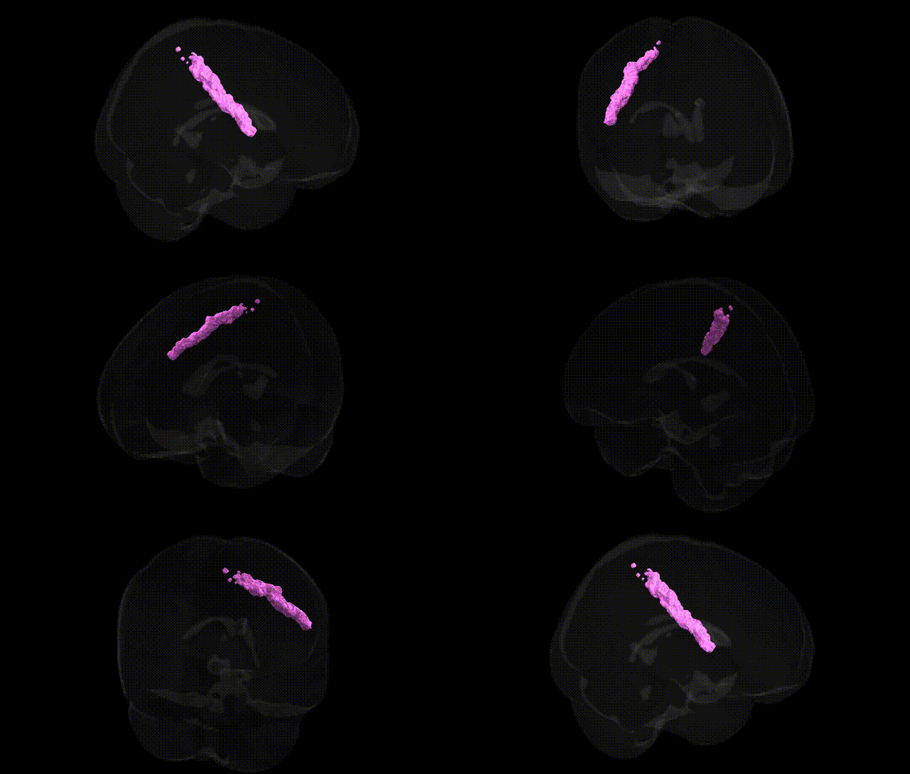
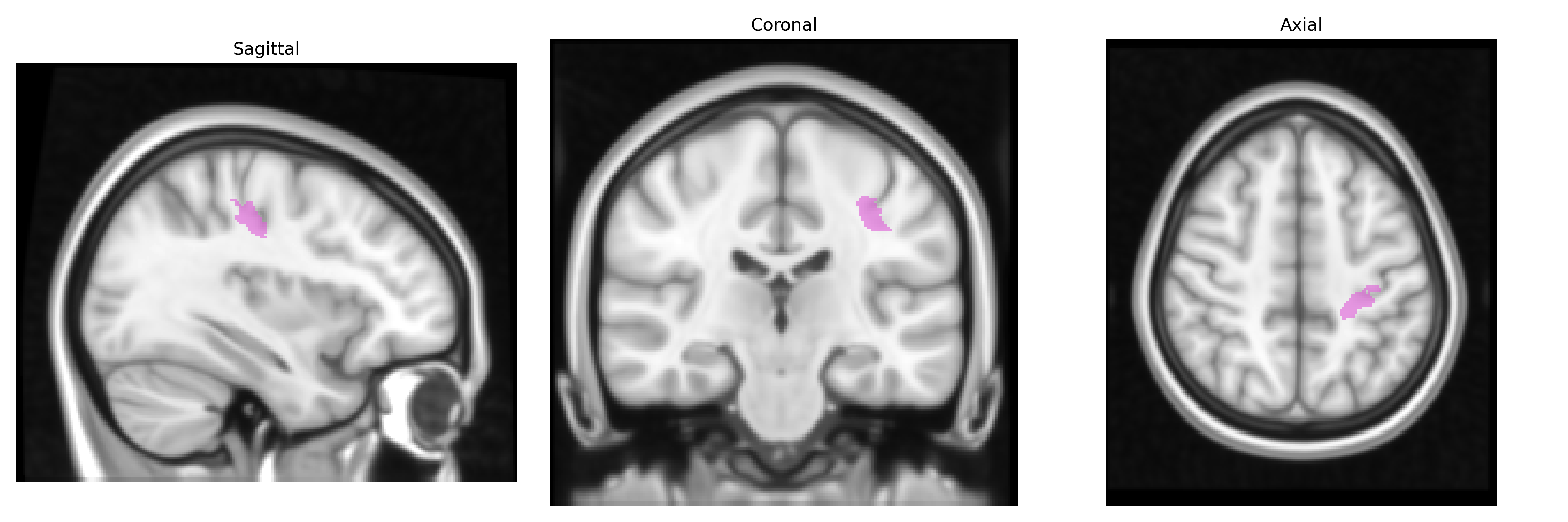
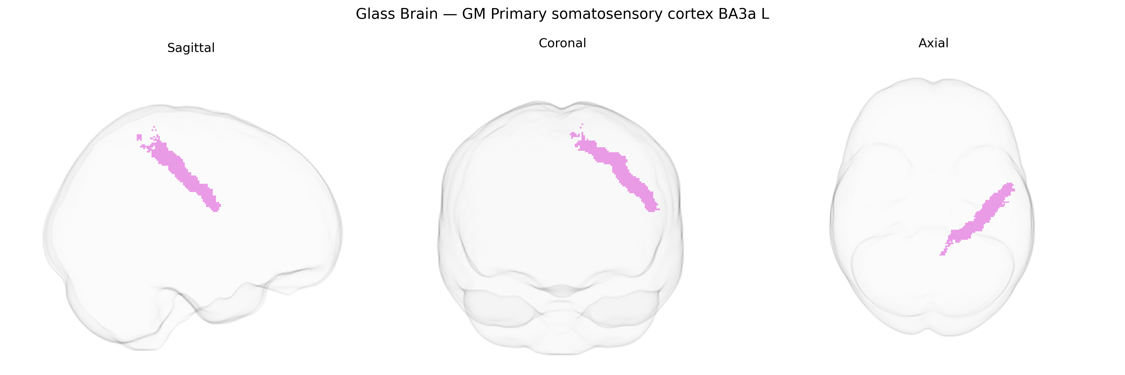
The bilateral GM Primary somatosensory cortex BA3a L, from the Juelich maxprob thr25 1mm Atlas, corresponds to the left-hemisphere portion of Brodmann area 3a within the primary somatosensory cortex, located in the anterior bank of the postcentral gyrus near the central sulcus. BA3a is primarily associated with processing proprioceptive and deep somatosensory information, receiving strong input from muscle spindles and joint receptors via thalamocortical projections. It contributes to the representation of body position and movement, forming part of the early cortical stages of the somatosensory processing hierarchy and interacting closely with adjacent areas 3b, 1, and 2 to integrate tactile and kinesthetic information. There is no direct Wikipedia article for BA3a; a related structure is the Primary somatosensory cortex.
The bilateral primary somatosensory cortex (S1) region encompassing Brodmann area 3a in the Juelich maxprob thr25 1 mm atlas has been implicated in genetic studies largely through its role in sensorimotor processing, pain perception, and neurodevelopmental traits, rather than through genes uniquely tied to this cytoarchitectonic parcel. GWAS and imaging-genetics studies have shown that individual differences in cortical thickness, surface area, and functional activation of S1—including BA3a-adjacent zones—are heritable and associated with common variants in genes involved in neurodevelopment (e.g., microtubule and axon guidance pathways), synaptic plasticity, and myelination, as well as with polygenic scores for general cognitive ability and educational attainment. Pain-related GWAS, including those on chronic pain, migraine, and neuropathic pain, frequently implicate somatosensory cortex activation and structure via imaging-derived phenotypes, though specific loci generally act through broad cortical or pain-network mechanisms rather than being unique to BA3a. Somatosensory cortex abnormalities, encompassing BA3a, have also been linked to polygenic risk for autism spectrum disorder and schizophrenia in imaging-genetics work, where genetic liability to these conditions correlates with altered S1 morphology or connectivity, and to motor or tremor-related phenotypes (e.g., essential tremor and dystonia) through studies of sensorimotor network genes affecting cerebello-thalamo-cortical circuits. Overall, genetic associations involving this region typically arise in the context of broader sensorimotor, pain, or neuropsychiatric networks, with BA3a contributing as part of a distributed heritable architecture rather than as a uniquely mapped genetic target.
Overview generated by GPT-4o (2026).
Region ID: 55
Hemisphere: bilateral
Atlas: Juelich maxprob thr25 1mm
Full Quality Version: Download MP4
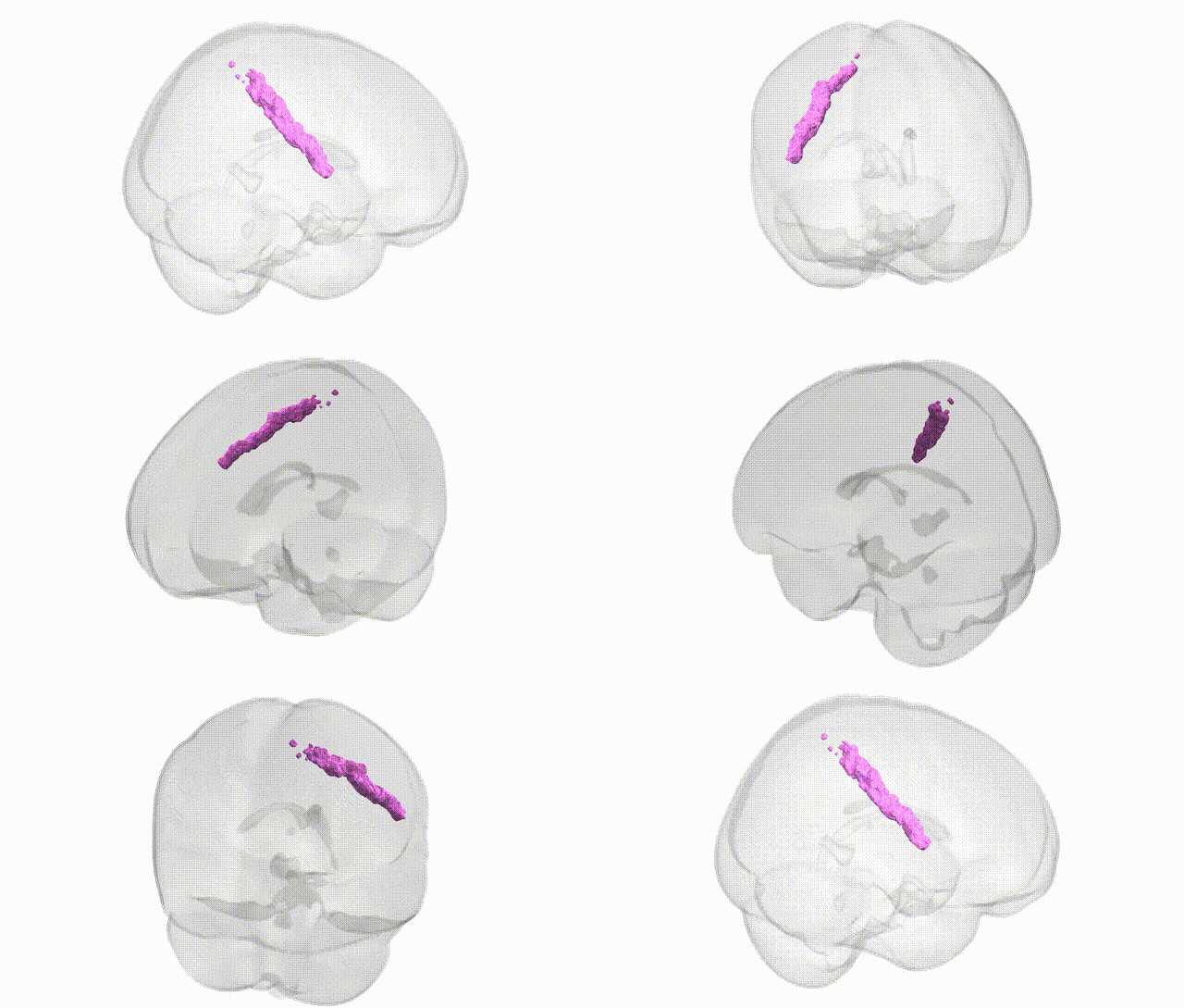
Full Quality Version: Download MP4
Wali Sidiqyar*, Gaurav Rudravaram*, Elyssa M. McMaster, Trent M. Schwartz, Adam M. Saunders, Kurt G. Schilling, Bennett A. Landman "Introducing SPINS: A Shared Public Visualization Library of Neuroanatomical Structures." Medical Imaging with Deep Learning- short paper
This resource is licensed under CC0 1.0 Universal (Public Domain).