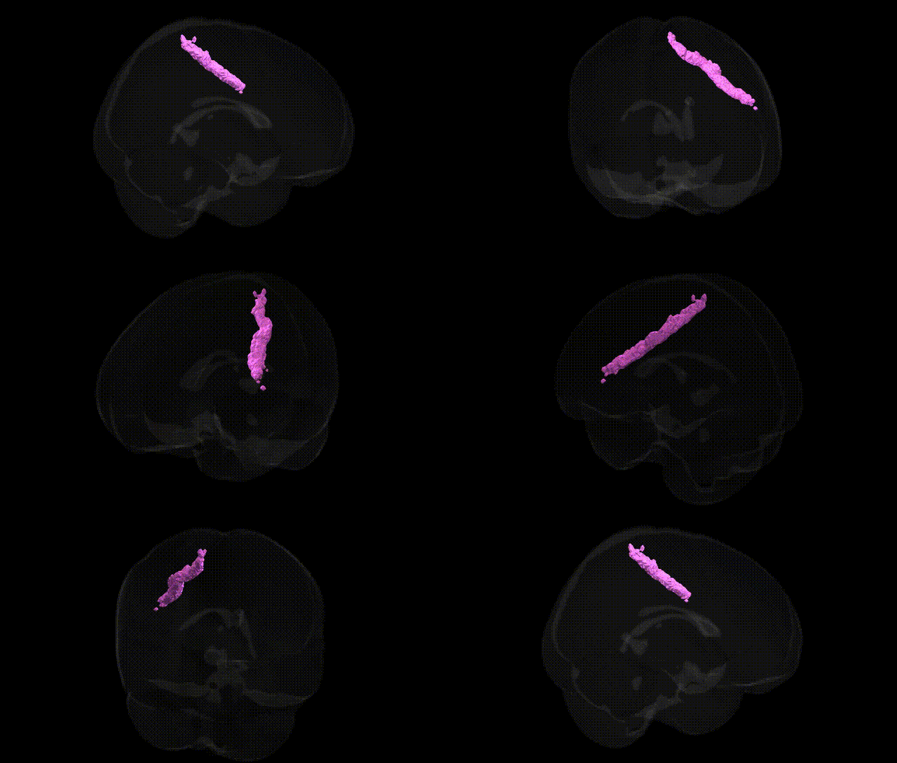
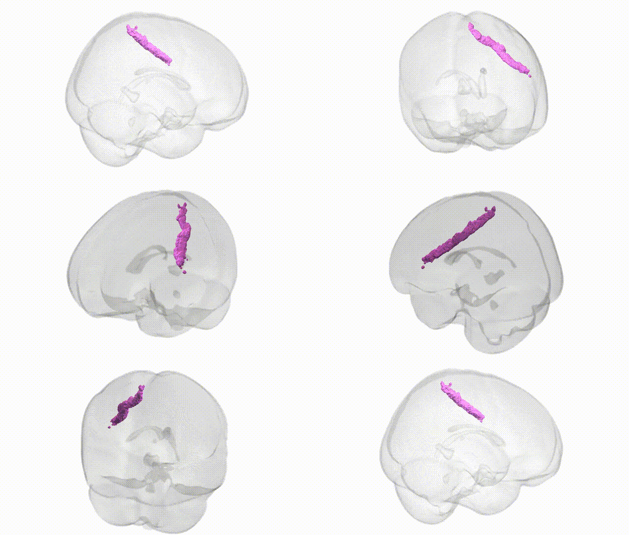
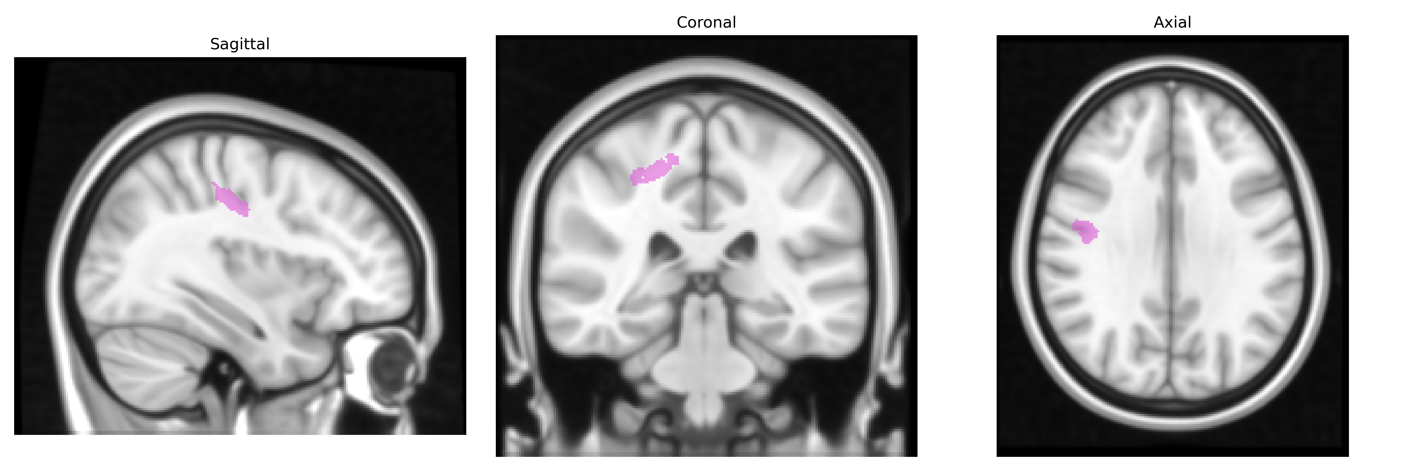
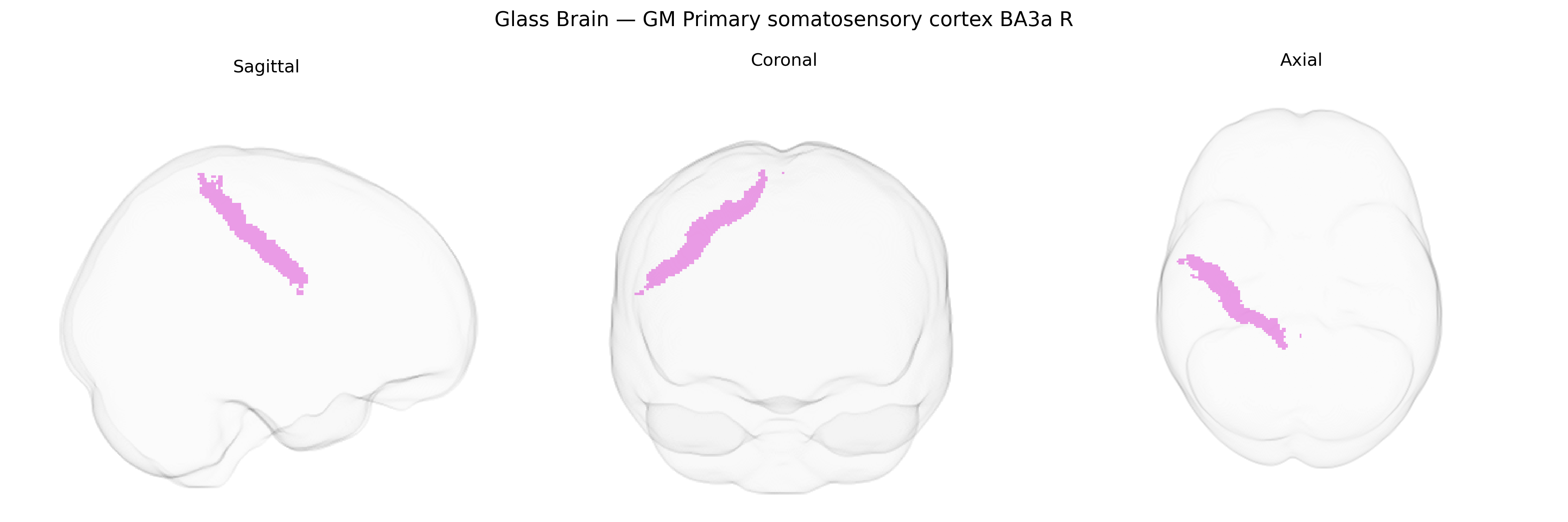
The bilateral GM Primary somatosensory cortex BA3a R, as defined in the Juelich maxprob thr25 1mm Atlas, corresponds to the granular field of Brodmann area 3a located in the posterior bank of the central sulcus within the postcentral gyrus, forming part of the primary somatosensory cortex. This region receives dense thalamocortical input from somatosensory relay nuclei and is particularly involved in processing proprioceptive information from muscle spindles and deep receptors, contributing to fine-grained representation of body position and movement. Neurons in BA3a exhibit a somatotopic organization and project to adjacent somatosensory fields (BA3b, BA1, BA2) as well as to motor-related areas, supporting sensorimotor integration required for precise motor control. There is no direct Wikipedia article for this specific atlas-defined region; a closely related structure is the Primary somatosensory cortex.
The bilateral GM primary somatosensory cortex (BA3a, right hemisphere) in the Juelich maxprob thr25 1 mm atlas has been implicated in several genetic and GWAS-based associations through imaging–genetics and neuropsychiatric studies that map variants to cortical thickness, surface area, and functional activity in the somatosensory strip. Common variants in genes affecting neurodevelopment and synaptic function (for example, BDNF, NRG1, and genes related to glutamatergic and GABAergic transmission) have been linked to structural variation in primary somatosensory areas, and large-scale cortical GWAS (e.g., ENIGMA and related consortia) report heritable influences on sensorimotor cortex morphology tied to loci involved in neuronal differentiation, axon guidance, and calcium signaling. Somatosensory cortex, including BA3a, appears in genetic studies of chronic pain and neuropathic pain, where polygenic risk for pain sensitivity and specific loci near genes such as SCN9A and CACNA1H are associated with altered somatosensory representation or responsiveness. Genetic risk for autism spectrum disorder and attention-deficit/hyperactivity disorder has been associated with atypical structure and connectivity in sensorimotor cortex, including BA3a-adjacent regions, through polygenic risk score–imaging analyses, implicating genes involved in synapse formation (e.g., SHANK family) and cortical circuit development. In schizophrenia and major depressive disorder, imaging–genetics work links polygenic risk to altered cortical thickness and functional activation in primary somatosensory and motor regions, suggesting that genetic architectures affecting widespread cortical networks encompass BA3a, though associations are typically broad and not specific to this subfield. Overall, current evidence indicates that BA3a participates in distributed genetic effects on cortical structure, pain processing, and neurodevelopmental/psychiatric liability, but no single gene or variant is uniquely or exclusively tied to the right BA3a field in available GWAS or atlas-based genetic analyses.
Overview generated by GPT-4o (2026).
Region ID: 56
Hemisphere: bilateral
Atlas: Juelich maxprob thr25 1mm
Full Quality Version: Download MP4
Full Quality Version: Download MP4
Wali Sidiqyar*, Gaurav Rudravaram*, Elyssa M. McMaster, Trent M. Schwartz, Adam M. Saunders, Kurt G. Schilling, Bennett A. Landman "Introducing SPINS: A Shared Public Visualization Library of Neuroanatomical Structures." Medical Imaging with Deep Learning- short paper
This resource is licensed under CC0 1.0 Universal (Public Domain).