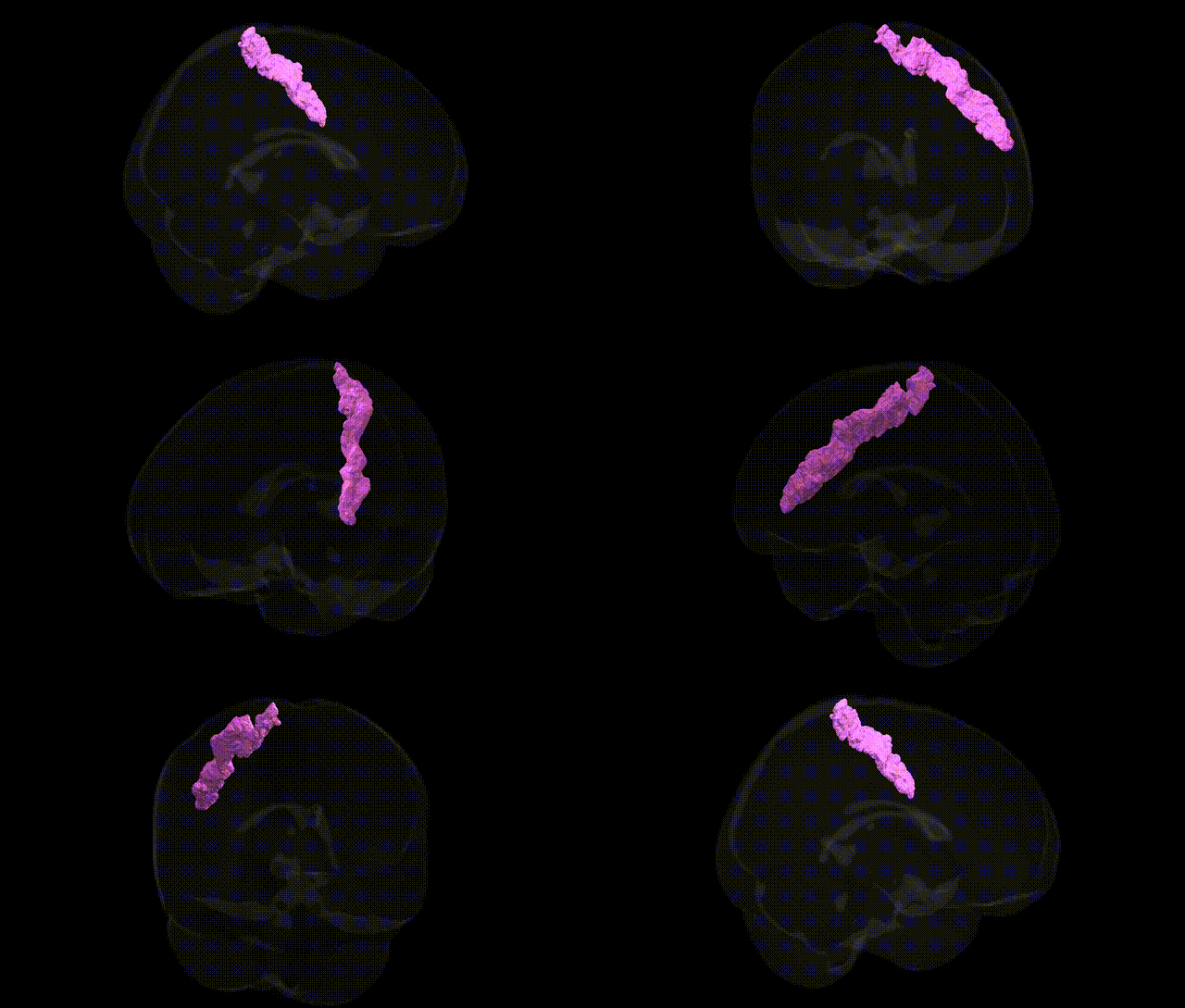
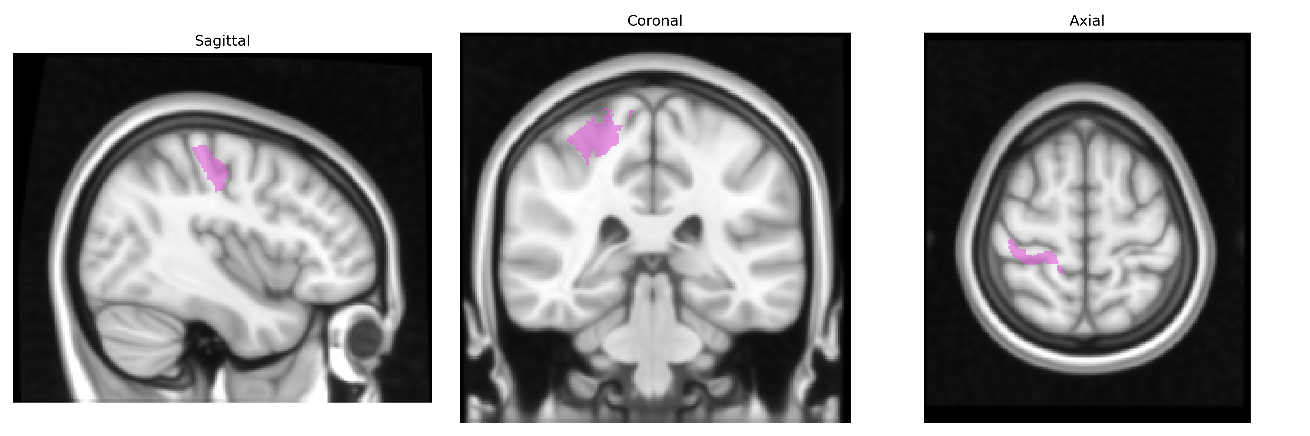
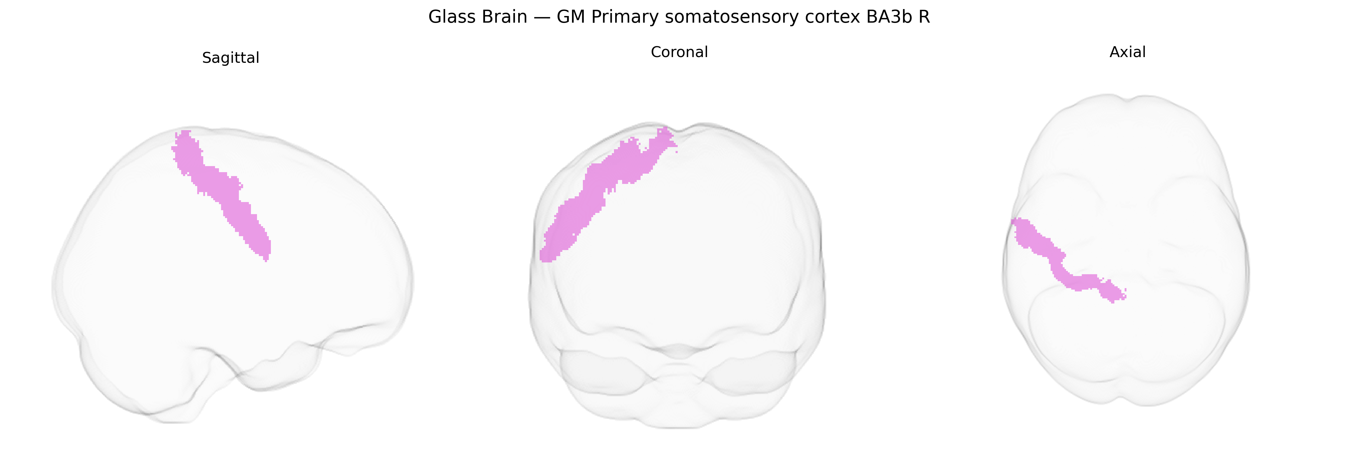
The bilateral GM Primary somatosensory cortex BA3b R (from the Juelich maxprob thr25 1mm Atlas) corresponds to Brodmann area 3b in the postcentral gyrus, a core subdivision of the primary somatosensory cortex responsible for processing cutaneous (tactile) input from the contralateral body. Neurons in BA3b receive dense thalamocortical projections from the ventral posterior nucleus of the thalamus and exhibit highly organized somatotopy, forming part of the primary somatosensory “homunculus.” This region is specialized for fine-grained discrimination of touch, including stimulus location, intensity, and texture, and serves as an initial cortical stage for tactile information that is further processed and integrated in adjacent somatosensory areas (BA1 and BA2). There is no direct Wikipedia article for “BA3b” as a standalone region; a closely related and encompassing structure is the Primary somatosensory cortex.
The bilateral gray matter primary somatosensory cortex BA3b (right hemisphere) from the Juelich maxprob atlas is a core node for tactile, proprioceptive, and pain processing, and genetic associations typically implicate broader somatosensory or sensorimotor networks rather than BA3b specifically. Large-scale imaging genetics studies (e.g., ENIGMA, UK Biobank) have linked common variants in genes related to synaptic function, neurodevelopment, and myelination (such as BDNF, GRIN2B, CNTNAP2, and multiple loci near microtubule and axon guidance genes) to interindividual differences in cortical thickness and surface area in primary sensorimotor regions, which encompass BA3b but do not isolate it. GWAS of chronic pain, tactile sensitivity, and neuropathic pain (including variants in SCN9A, SCN10A, CACNA2D1, and genes influencing peripheral nociception and central pain modulation) have shown functional and structural correlates in primary somatosensory cortex, suggesting genetically influenced vulnerability of BA3b-related circuits in pain disorders, though the associations are again regionally broad. Neurodevelopmental and neuropsychiatric GWAS—for autism spectrum disorder, ADHD, schizophrenia, and dyslexia—have repeatedly implicated genes involved in cortical patterning and synaptic plasticity (e.g., FOXP1/FOXP2 pathways, NRXN1, SHANK3, and others), with downstream imaging findings of altered somatosensory and sensorimotor cortex structure and connectivity, consistent with BA3b involvement but without fine-grained, atlas-specific genetic mapping. Overall, current genetic evidence points to polygenic influences on the structure and function of the primary somatosensory cortex as part of distributed sensorimotor networks, with no robust GWAS identifying variants uniquely associated with the Juelich-defined BA3b R region, and most links to disorders or traits are inferred from broader cortical and pain-related phenotypes rather than direct locus-to-BA3b associations.
Overview generated by GPT-4o (2026).
Region ID: 58
Hemisphere: bilateral
Atlas: Juelich maxprob thr25 1mm
Full Quality Version: Download MP4
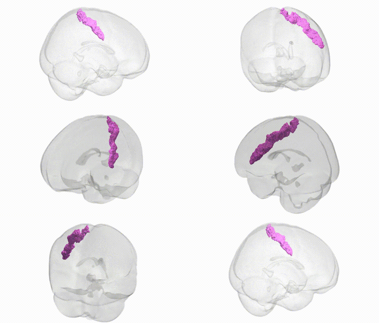
Full Quality Version: Download MP4
Wali Sidiqyar*, Gaurav Rudravaram*, Elyssa M. McMaster, Trent M. Schwartz, Adam M. Saunders, Kurt G. Schilling, Bennett A. Landman "Introducing SPINS: A Shared Public Visualization Library of Neuroanatomical Structures." Medical Imaging with Deep Learning- short paper
This resource is licensed under CC0 1.0 Universal (Public Domain).