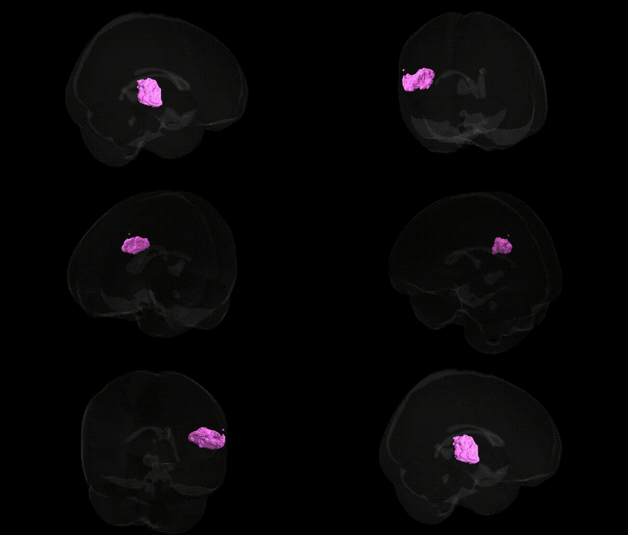
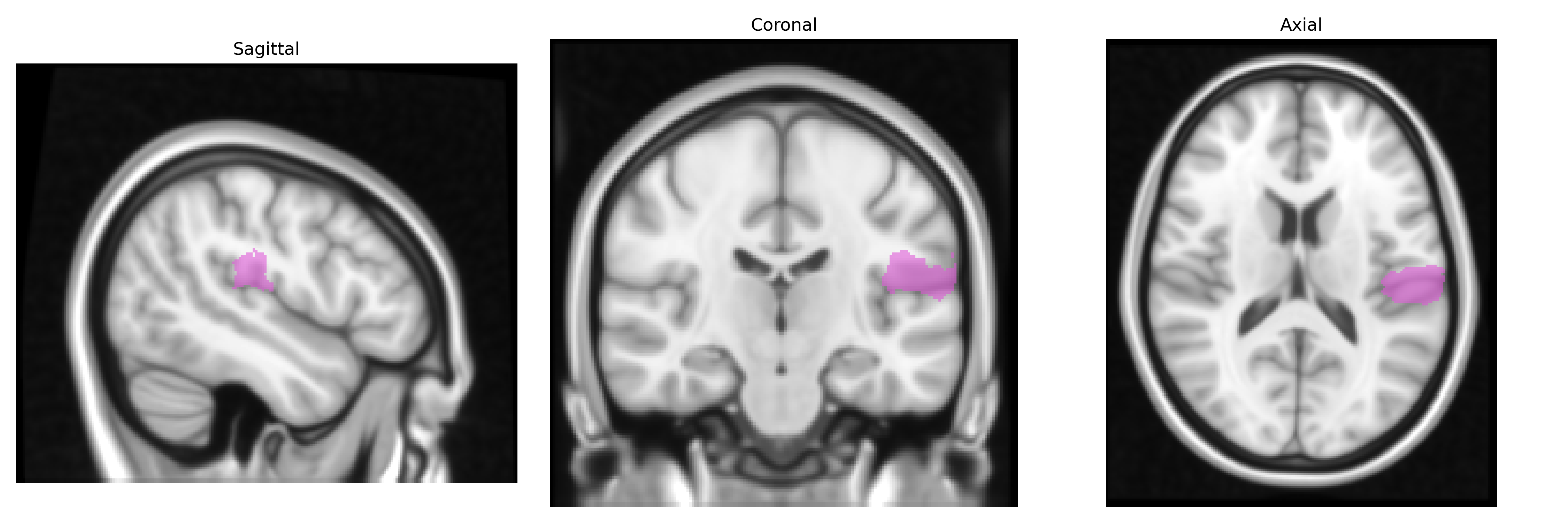
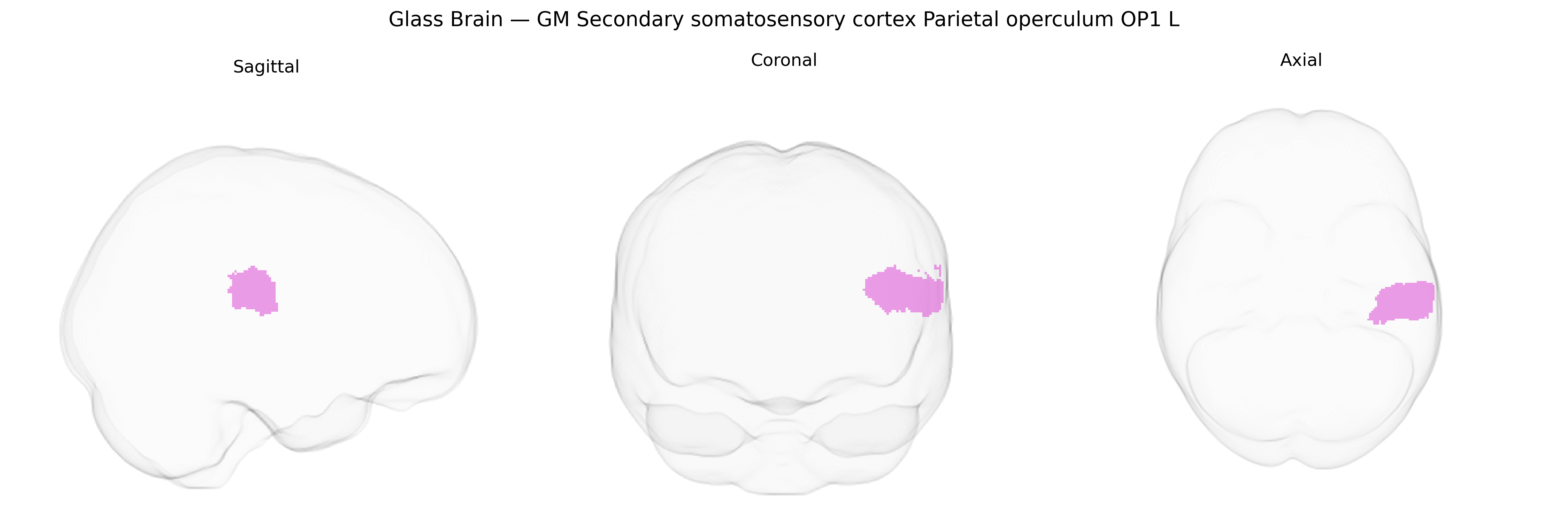
The bilateral GM Secondary somatosensory cortex Parietal operculum OP1 L, as defined in the Juelich maxprob thr25 1 mm atlas, corresponds to a cytoarchitectonically distinct portion of the human secondary somatosensory cortex (S2) located within the parietal operculum overlying the insula. This region participates in higher-order somatosensory processing, including integration of tactile, nociceptive, and proprioceptive inputs, bilateral representation of the body, and contributions to sensorimotor integration and tactile object recognition. Neurons within OP1 exhibit multimodal convergence and play a role in perceptual functions such as texture discrimination, somatosensory memory, and possibly aspects of pain perception and body schema. OP1 is reciprocally connected with primary somatosensory cortex, other opercular fields, insular cortex, and thalamic nuclei, forming part of a distributed somatosensory network. There is no direct Wikipedia article for OP1; a related structure is the Secondary somatosensory cortex.
The bilateral GM Secondary somatosensory cortex Parietal operculum OP1 L (Juelich maxprob thr25 1mm Atlas) is part of the human SII/opercular somatosensory system, and genetic associations involving this region are largely inferred from imaging genetics and GWAS of cortical structure and pain-related traits rather than from studies targeting OP1 specifically. Large-scale neuroimaging GWAS (e.g., ENIGMA, UK Biobank) have identified common variants in genes involved in neurodevelopment, synaptic function, and axon guidance—such as BDNF, microtubule-associated and cell-adhesion genes, and loci in neurodevelopmental pathways—associated with thickness, surface area, or volume in lateral parietal and opercular cortices, indirectly implicating OP1. Functionally, the parietal operculum/SII region has been linked to pain perception, tactile processing, and interoception, and candidate-gene as well as GWAS-based imaging studies in chronic pain conditions (e.g., neuropathic pain, migraine, fibromyalgia) and somatosensory disorders have reported associations with polymorphisms in COMT, OPRM1, and serotonergic and dopaminergic genes that modulate activity or structural integrity in opercular somatosensory areas. Additionally, risk variants for neurodevelopmental and psychiatric conditions—such as schizophrenia, autism spectrum disorder, and ADHD—identified in large GWAS (e.g., CACNA1C, CNTNAP2, and other synaptic/neurodevelopmental loci) have been related to altered activation or morphology in the parietal operculum and adjacent insulo-opercular networks during tactile, pain, or multisensory tasks, suggesting that genetic influences on these disorders partially act through circuits encompassing OP1, though no single gene has been established as specifically or exclusively associated with this atlas-defined subregion.
Overview generated by GPT-4o (2026).
Region ID: 59
Hemisphere: bilateral
Atlas: Juelich maxprob thr25 1mm
Full Quality Version: Download MP4
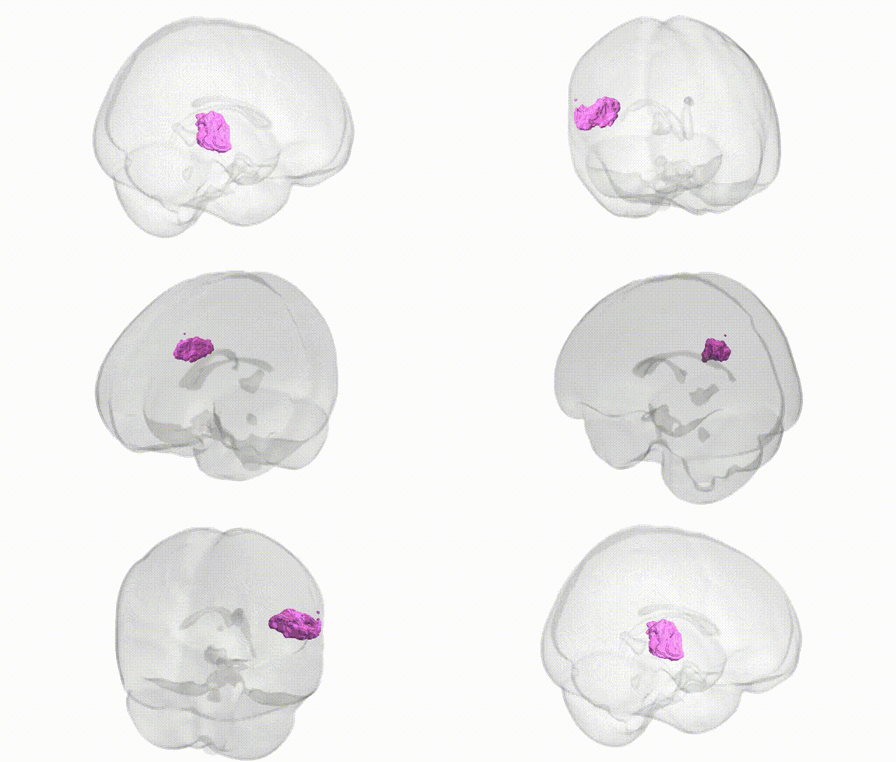
Full Quality Version: Download MP4
Wali Sidiqyar*, Gaurav Rudravaram*, Elyssa M. McMaster, Trent M. Schwartz, Adam M. Saunders, Kurt G. Schilling, Bennett A. Landman "Introducing SPINS: A Shared Public Visualization Library of Neuroanatomical Structures." Medical Imaging with Deep Learning- short paper
This resource is licensed under CC0 1.0 Universal (Public Domain).