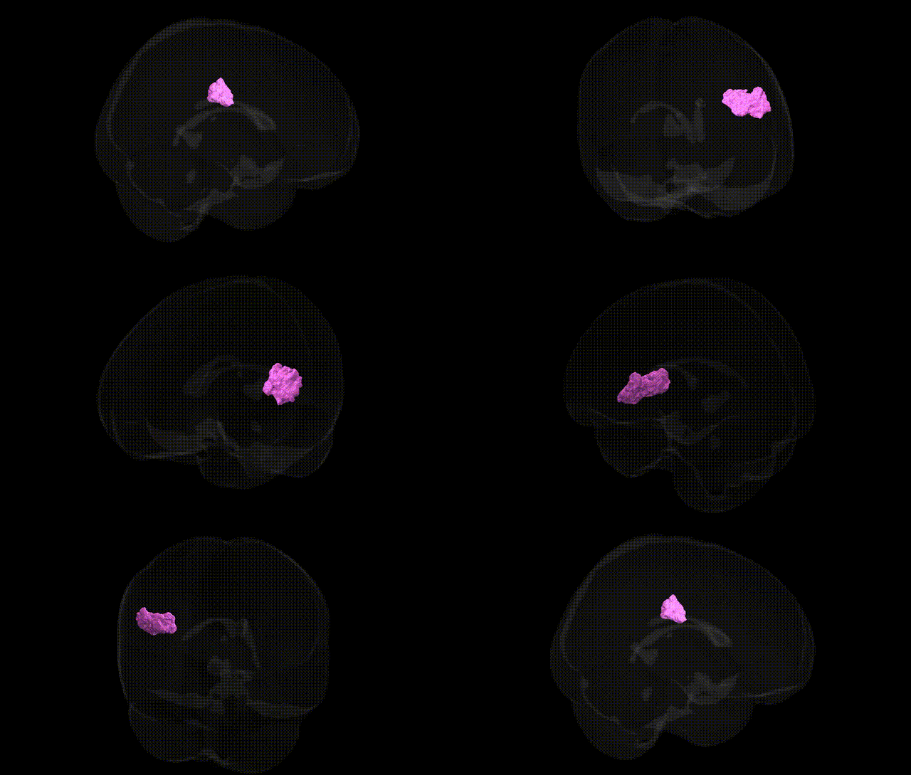
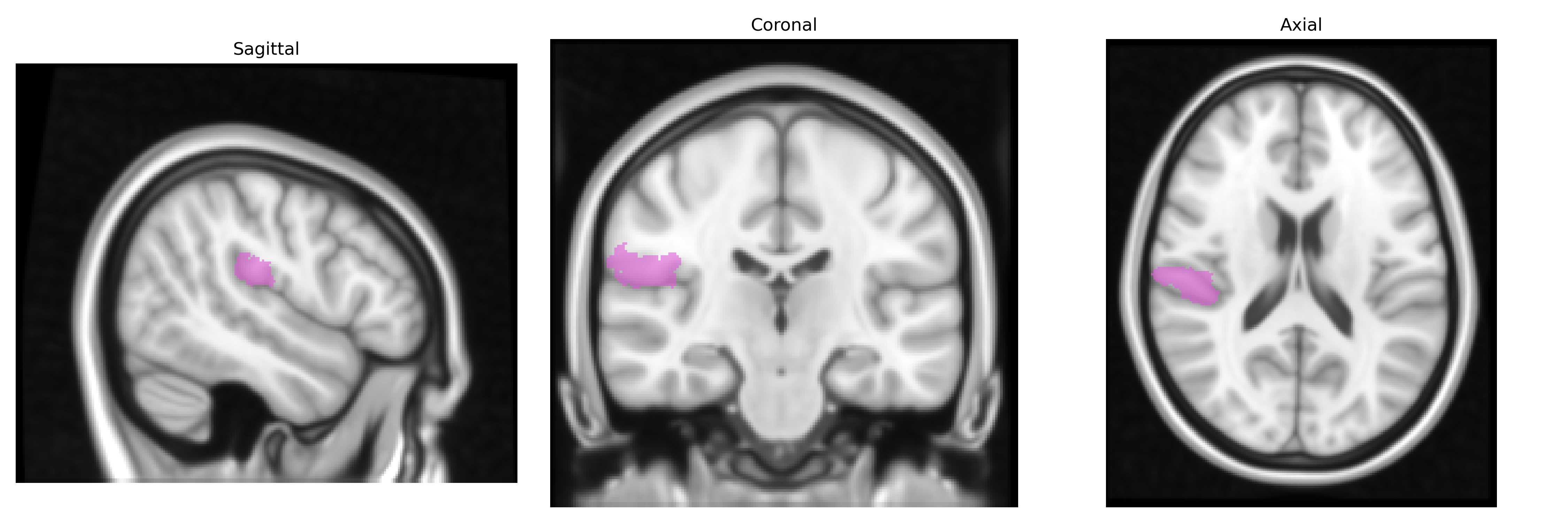
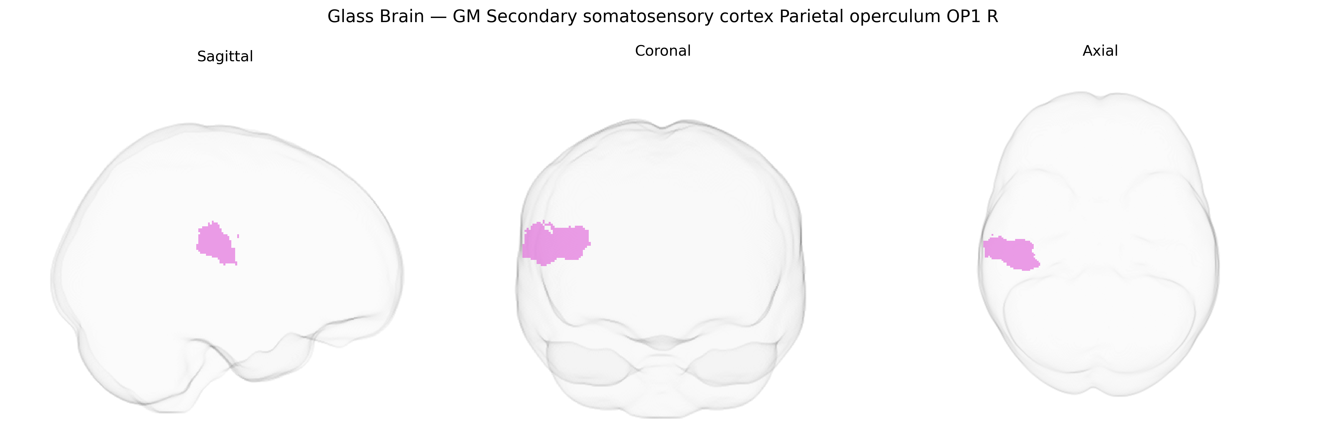
The bilateral GM Secondary somatosensory cortex Parietal operculum OP1 R, as defined in the Juelich maxprob thr25 1mm Atlas, corresponds to a cytoarchitectonically distinct subregion of the human secondary somatosensory cortex (SII) located within the parietal operculum of the Sylvian fissure. OP1 is involved in higher-order somatosensory processing, including integration of tactile, proprioceptive, and nociceptive information, and contributes to functions such as texture and shape discrimination, body perception, and aspects of sensorimotor integration. This region receives input from primary somatosensory cortex (S1) and thalamic nuclei and projects to multisensory and motor-related areas, supporting complex perceptual and cognitive aspects of touch. There is no direct Wikipedia article for OP1; a related structure is the Parietal operculum.
The bilateral GM Secondary somatosensory cortex Parietal operculum OP1 R, as defined in the Juelich maxprob thr25 1 mm atlas, corresponds largely to posterior insula–parietal opercular regions implicated in somatosensory, pain, and sensorimotor integration, and genetic associations to this area arise mainly from imaging–genetics and GWAS of cortical morphology, somatosensory function, and pain-related traits. Large-scale brain-structure GWAS (e.g., ENIGMA and UK Biobank–based studies) have identified common variants in loci such as 17q21.31 (encompassing MAPT/KANSL1), 3p24–p25, and 6q22–q23 that influence cortical thickness or surface area of perisylvian/insular–parietal opercular regions overlapping OP1, while polygenic scores for neuroticism, educational attainment, and general cognitive ability show modest associations with structural variation in adjacent secondary somatosensory and insular cortices. Functional imaging–genetics work links polymorphisms in COMT (Val158Met), OPRM1 (A118G), and GCH1 to altered pain-evoked activation in secondary somatosensory and parietal opercular cortex, and rare-variant or CNV studies in neurodevelopmental and epilepsy cohorts (e.g., 22q11.2 deletions, SCN1A and GRIN2A mutations) frequently report abnormalities involving perisylvian–opercular networks that include OP1. Psychiatric and neurological GWAS for disorders such as schizophrenia, major depression, autism spectrum disorder, and chronic pain indicate that their polygenic risk scores correlate with patterns of cortical thinning or altered gyrification in somatosensory–insular–opercular areas, although associations to OP1 specifically are generally indirect and mediated through distributed networks rather than single-region, single-gene effects.
Overview generated by GPT-4o (2026).
Region ID: 60
Hemisphere: bilateral
Atlas: Juelich maxprob thr25 1mm
Full Quality Version: Download MP4
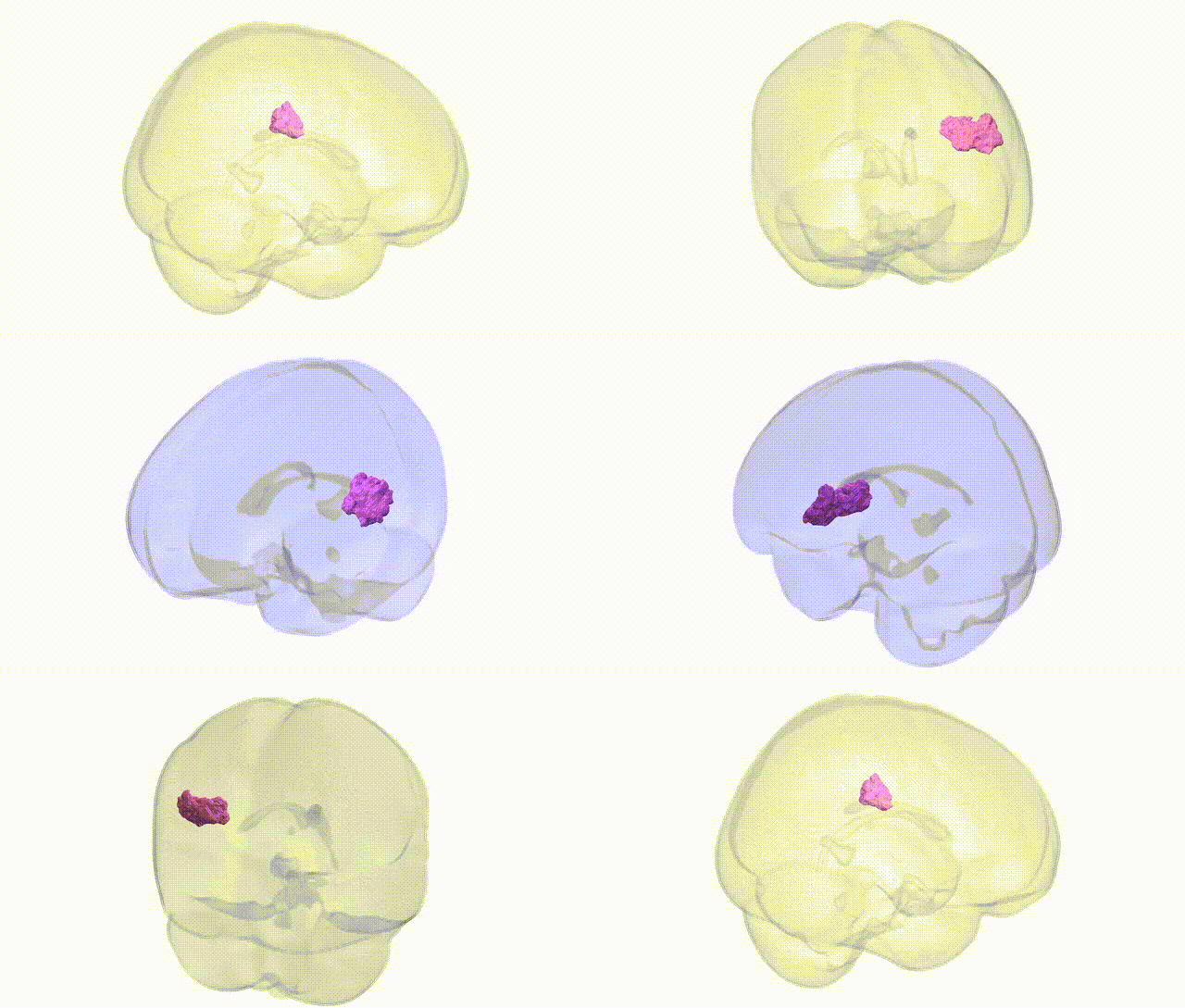
Full Quality Version: Download MP4
Wali Sidiqyar*, Gaurav Rudravaram*, Elyssa M. McMaster, Trent M. Schwartz, Adam M. Saunders, Kurt G. Schilling, Bennett A. Landman "Introducing SPINS: A Shared Public Visualization Library of Neuroanatomical Structures." Medical Imaging with Deep Learning- short paper
This resource is licensed under CC0 1.0 Universal (Public Domain).