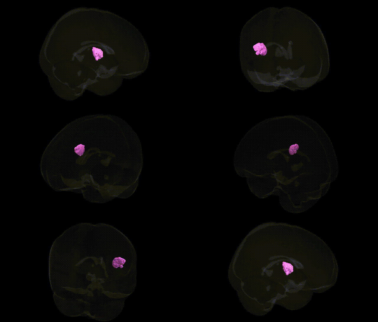
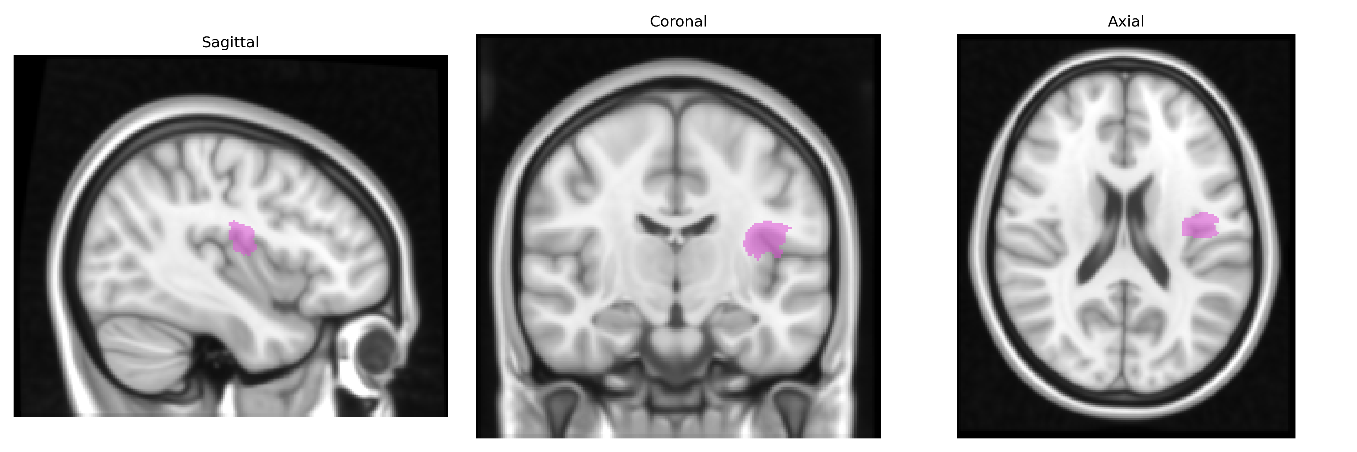
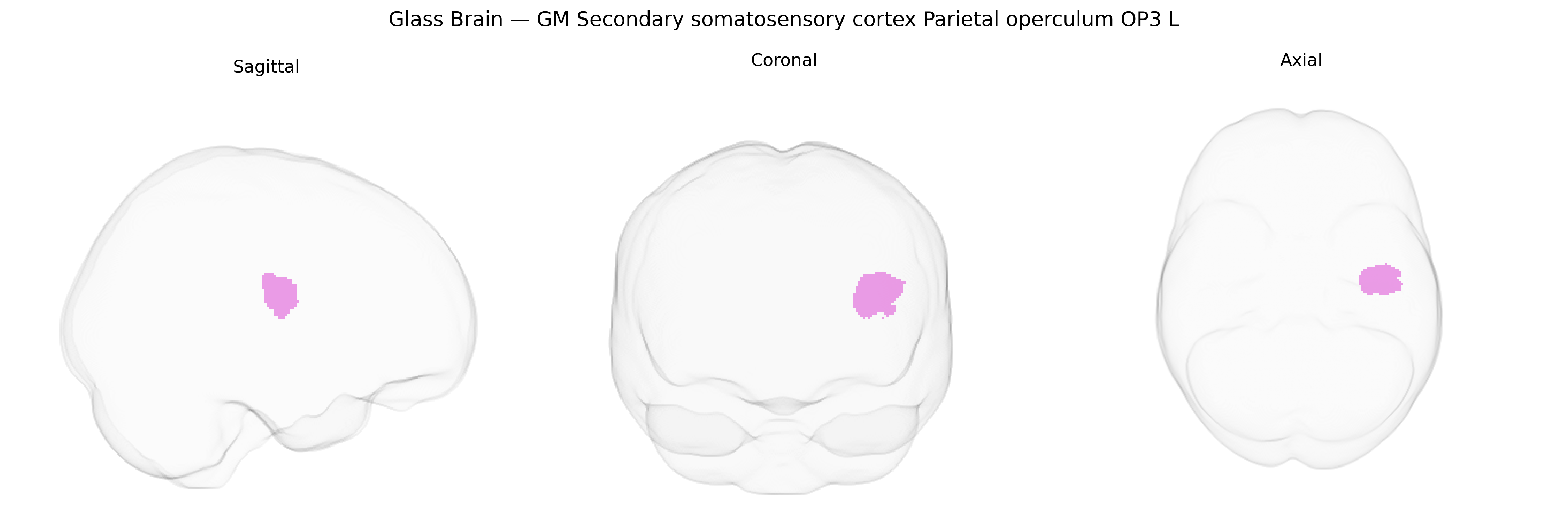
The bilateral GM Secondary somatosensory cortex Parietal operculum OP3 L, as defined in the Juelich maxprob thr25 1mm atlas, corresponds to a cytoarchitectonically distinct subdivision of the human parietal operculum that forms part of the secondary somatosensory cortex (SII). This region receives convergent somatosensory input from primary somatosensory cortex and thalamic nuclei and is implicated in higher-order processing of tactile, nociceptive, and proprioceptive information, including texture discrimination, bilateral integration of somatosensory signals, and aspects of body perception and sensorimotor integration. OP3 exhibits strong connectivity with other opercular regions, insular cortex, and sensorimotor networks, thereby contributing to multimodal integration and the representation of complex somatic sensations. There is no direct link for OP3; a related structure is the Parietal operculum.
The bilateral GM secondary somatosensory cortex (parietal operculum OP3 L) in the Juelich maxprob atlas is part of the broader somatosensory and opercular network, and genetic associations typically emerge from imaging genetics and GWAS of cortical structure or function rather than OP3-specific studies. Large-scale GWAS of cortical thickness and surface area (e.g., ENIGMA and UK Biobank cohorts) have identified variants in genes such as CENPW, TBR1, and NRXN1 that influence parietal and opercular morphology, though these findings generally map to regional parcellations (somatosensory, parietal, or insular cortices) rather than the OP3 field alone. Functional imaging genetics studies link common polymorphisms in dopaminergic and glutamatergic genes (e.g., COMT, DRD2, GRIN2B) to altered activation in somatosensory and pain-processing cortices, including parietal operculum, during nociceptive, tactile, and sensorimotor tasks. GWAS and candidate gene studies in chronic pain, migraine, and neuropathic pain implicate genes involved in neuroinflammation (e.g., TNF, IL6), ion channel function (SCN9A, CACNA1A), and synaptic plasticity (BDNF) whose carriers show altered somatosensory cortex structure or reactivity, with parietal operculum frequently appearing in imaging endophenotypes. Additional associations arise from GWAS of traits such as somatosensory processing, pain sensitivity, and touch perception, where risk variants modulate activity in secondary somatosensory cortex–parietal operculum circuits, and from neuropsychiatric GWAS (e.g., autism spectrum disorder, schizophrenia, and anxiety-related traits) that report secondary somatosensory and opercular changes as intermediate phenotypes, suggesting that polygenic influences on sensory integration and interoception partly converge on this region, although no major study has yet isolated OP3 L as a uniquely and reproducibly associated genetic locus.
Overview generated by GPT-4o (2026).
Region ID: 63
Hemisphere: bilateral
Atlas: Juelich maxprob thr25 1mm
Full Quality Version: Download MP4
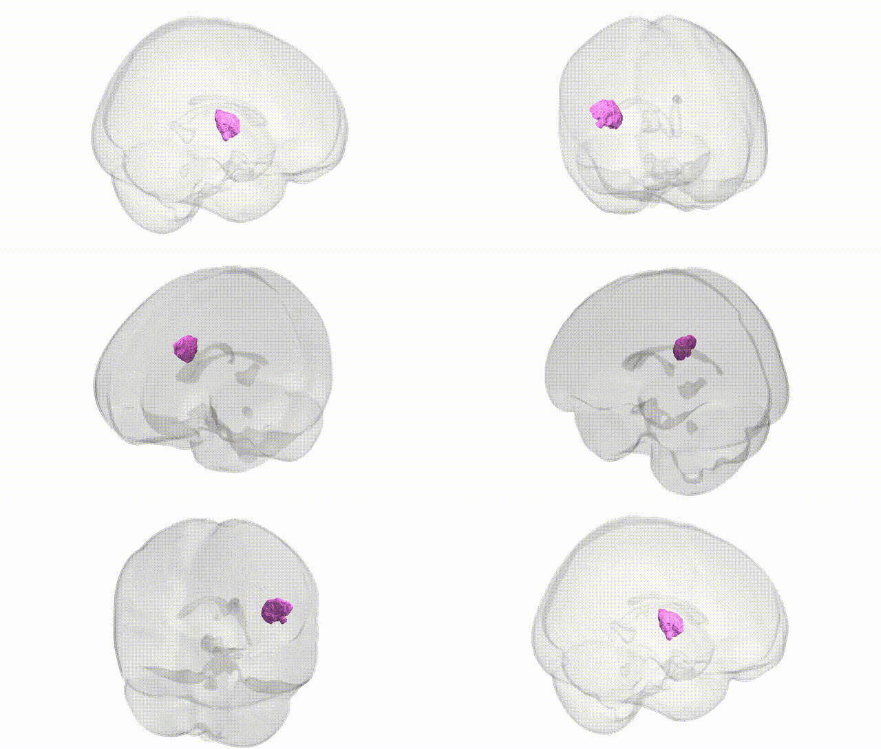
Full Quality Version: Download MP4
Wali Sidiqyar*, Gaurav Rudravaram*, Elyssa M. McMaster, Trent M. Schwartz, Adam M. Saunders, Kurt G. Schilling, Bennett A. Landman "Introducing SPINS: A Shared Public Visualization Library of Neuroanatomical Structures." Medical Imaging with Deep Learning- short paper
This resource is licensed under CC0 1.0 Universal (Public Domain).