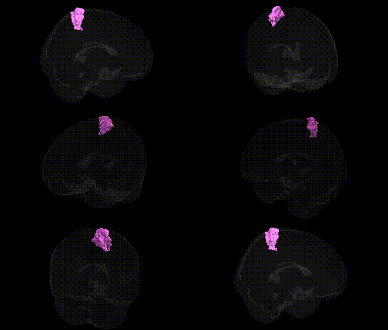
The bilateral GM Superior parietal lobule 5L L region, as defined in the Juelich maxprob thr25 1 mm atlas, corresponds to a left-hemispheric subregion of Brodmann area 5 located within the superior parietal lobule, posterior to the primary somatosensory cortex (areas 3, 1, and 2) and anterior to the precuneus. This area participates in higher-order somatosensory processing and sensorimotor integration, receiving multimodal input (tactile, proprioceptive, and visual) and contributing to body schema, spatial orientation, and the guidance of limb movements in peripersonal space. Functionally, it is implicated in coordinating reaching and grasping, integrating sensory feedback with motor plans, and supporting aspects of visuomotor transformation and attention to spatial location. There is no direct Wikipedia article for “Area 5L”; a related structure in which it is embedded is the Superior parietal lobule.
The bilateral GM Superior parietal lobule 5L L region (Juelich maxprob thr25 1mm Atlas) lies within the superior parietal lobule (SPL), a hub for visuospatial integration, attention, and sensorimotor coordination, and genetic associations are typically reported at the level of SPL or parietal cortex rather than the specific 5L subfield. Large-scale neuroimaging–genetics studies (e.g., ENIGMA, UK Biobank) have identified common variants near genes such as HMGA2, IGF1, WNT signaling components, and microtubule- or synapse-related genes that influence parietal cortical thickness and surface area, including superior parietal regions; polygenic architecture shows overlap with general cognitive ability and educational attainment. GWAS of cortical morphology consistently implicate superior parietal regions in traits such as intelligence, working memory, and visuospatial performance, with shared genetic variance between SPL structure and these cognitive traits. Parietal cortex volume and function, encompassing superior parietal areas, have been linked to risk loci for neurodevelopmental and psychiatric conditions, including ADHD, schizophrenia, and autism spectrum disorder, where genes affecting synaptic plasticity, neuronal migration, and cortical patterning (e.g., CACNA1C, CNTNAP2, MIR137-related loci) contribute to altered parietal morphometry or connectivity. In neurodegenerative disease, genetic risk for Alzheimer’s disease (e.g., APOE ε4, CLU, PICALM) and posterior cortical atrophy shows effects on posterior parietal regions, including superior parietal cortex, which exhibits early atrophy and hypometabolism in carriers of high-risk alleles. While no robust GWAS signals have been reported specifically for the 5L L subregion, convergent evidence indicates that the superior parietal lobule is a genetically influenced node whose structure and function mediate the impact of polygenic risk for cognition, attentional control, and several neuropsychiatric and neurodegenerative disorders.
Overview generated by GPT-4o (2026).
Region ID: 69
Hemisphere: bilateral
Atlas: Juelich maxprob thr25 1mm
Full Quality Version: Download MP4
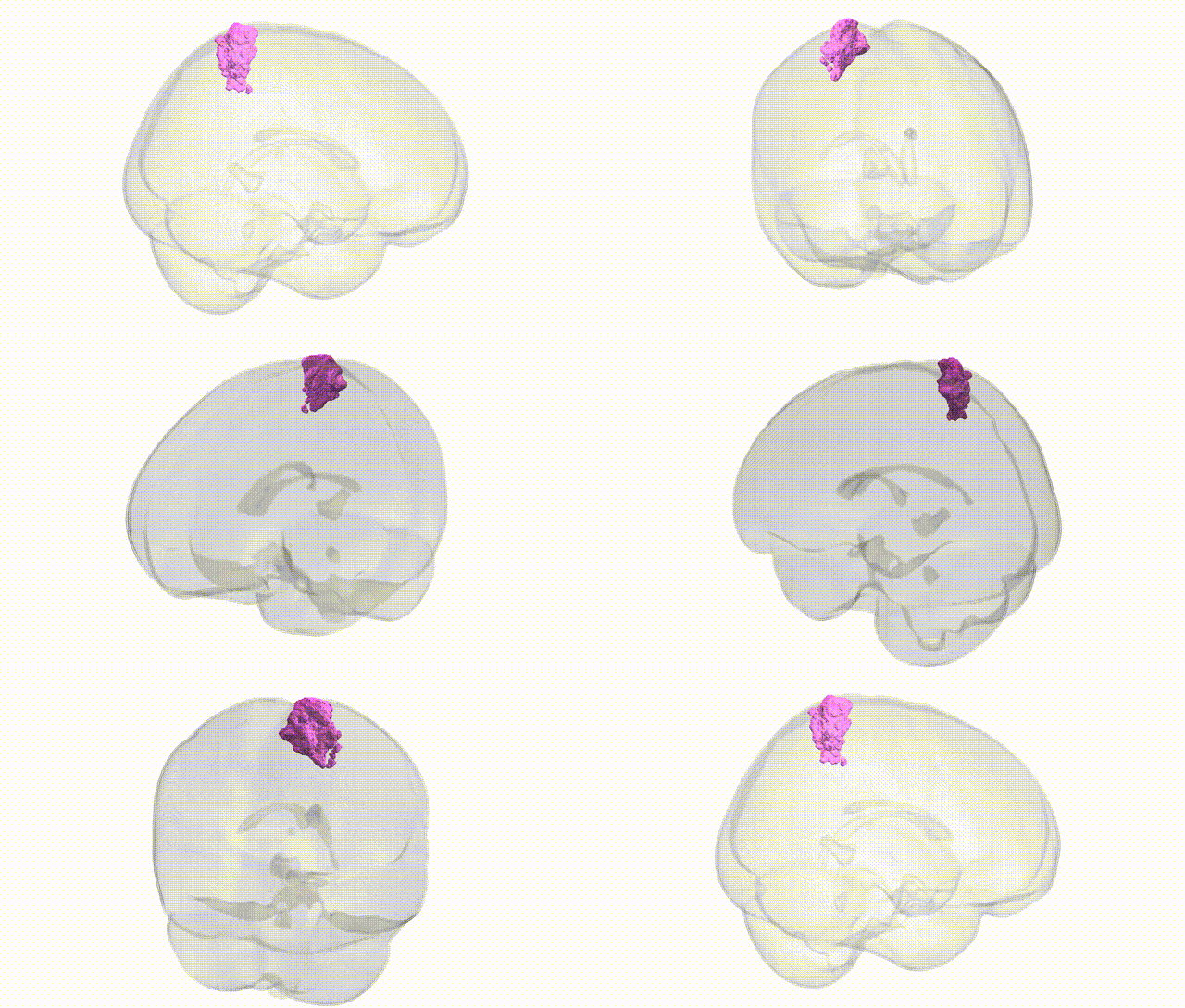
Full Quality Version: Download MP4
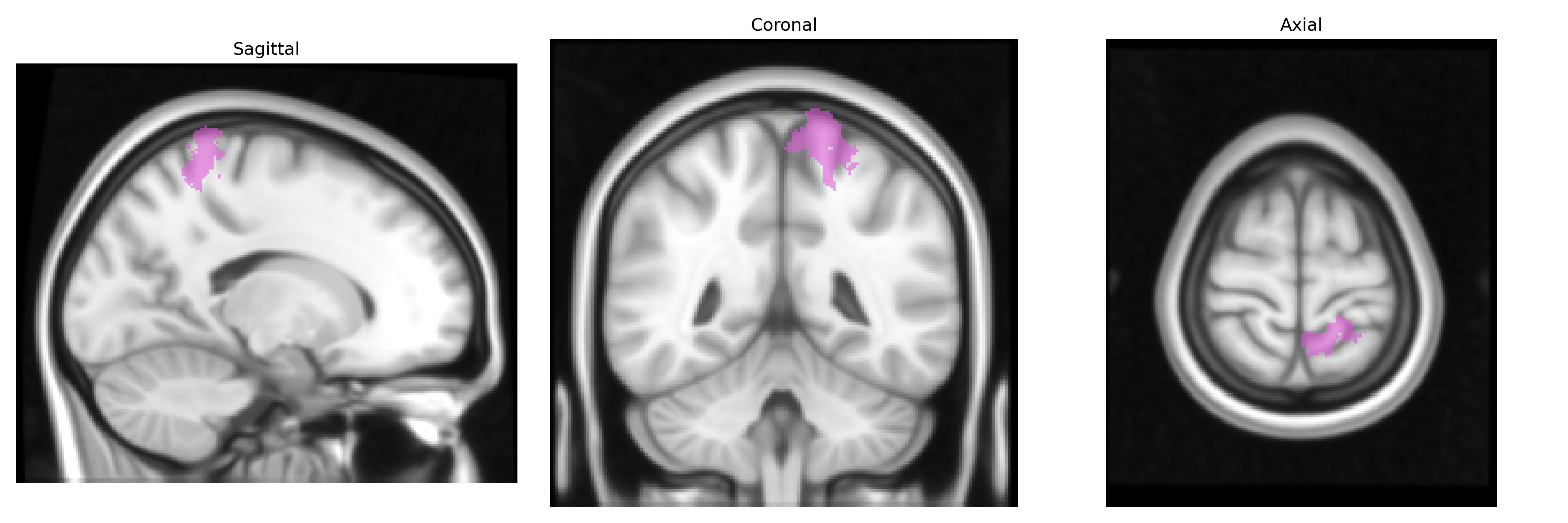
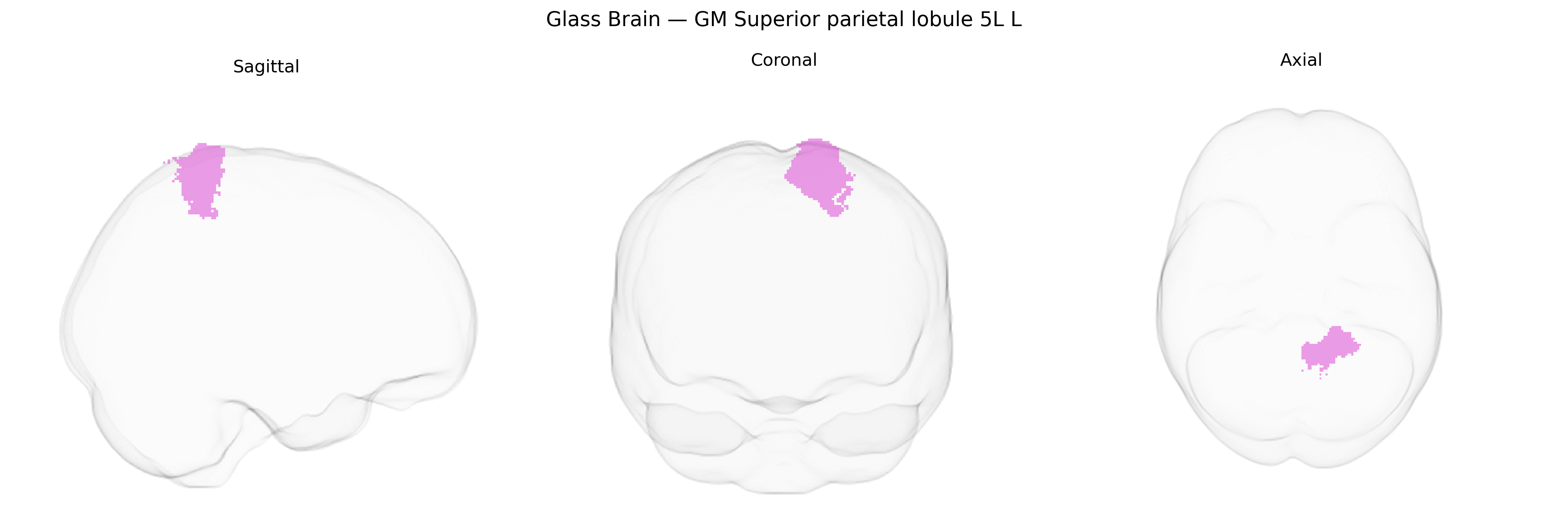
Wali Sidiqyar*, Gaurav Rudravaram*, Elyssa M. McMaster, Trent M. Schwartz, Adam M. Saunders, Kurt G. Schilling, Bennett A. Landman "Introducing SPINS: A Shared Public Visualization Library of Neuroanatomical Structures." Medical Imaging with Deep Learning- short paper
This resource is licensed under CC0 1.0 Universal (Public Domain).