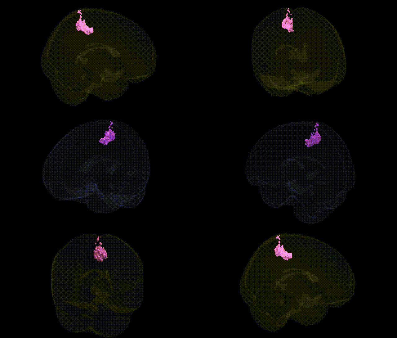
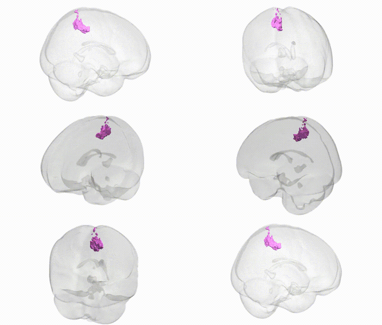
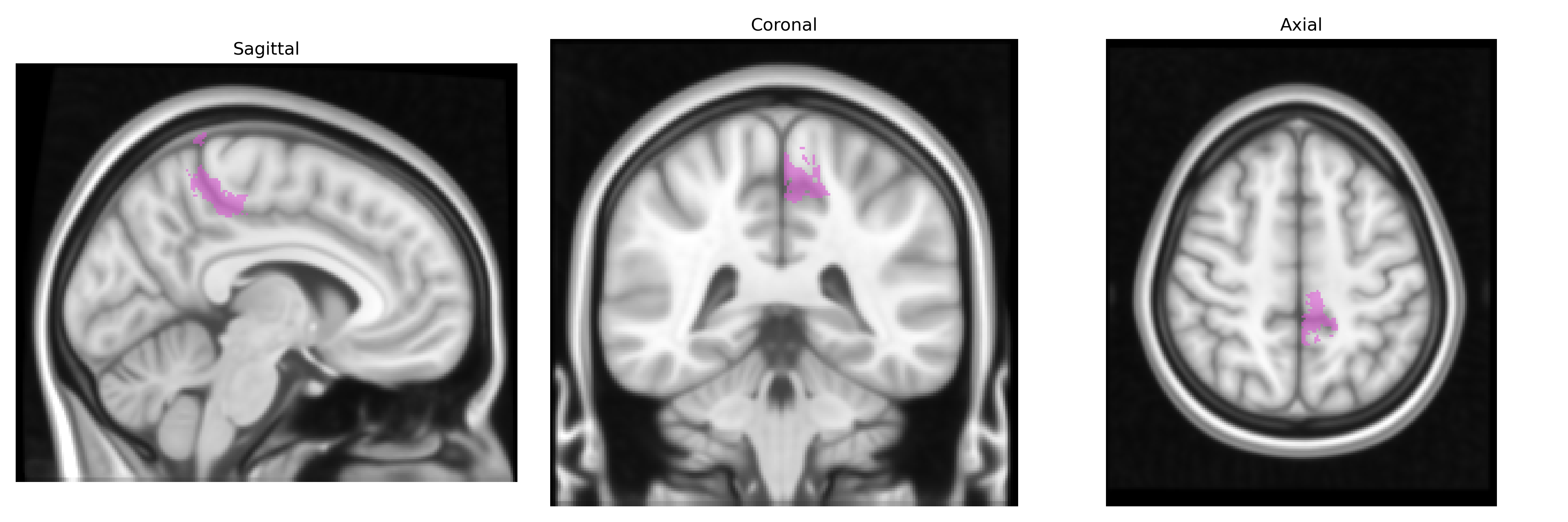
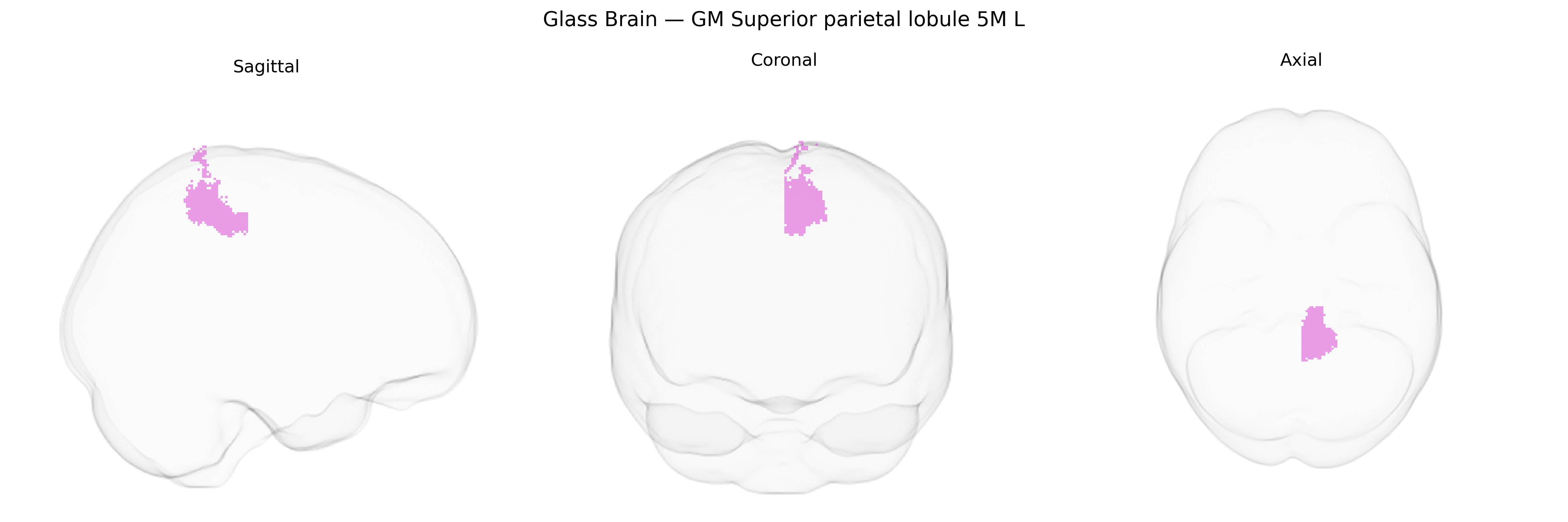
The bilateral GM Superior parietal lobule area 5M (L) from the Juelich maxprob thr25 1mm atlas corresponds to a medial subdivision of Brodmann area 5 located in the superior parietal lobule, bordering the precuneus and adjacent superior parietal fields (e.g., areas 7A/7P). This region is part of the posterior parietal cortex and is implicated in higher-order somatosensory integration, particularly the transformation of proprioceptive and tactile input into sensorimotor representations that support limb coordination, body schema, and visually guided actions. Neuronal populations in 5M exhibit multimodal responses, integrating somatic, visual, and sometimes vestibular signals, thereby contributing to the planning and online control of reaching and grasping movements as well as spatial attention within peripersonal space. There is no direct link for “Superior parietal lobule 5M” on Wikipedia; a related structure within which this area is located is the Superior parietal lobule.
The bilateral GM Superior parietal lobule 5M L, a dorsal parietal subregion implicated in visuospatial integration, attention, and sensorimotor coordination, has been indirectly linked to several genetic influences through imaging genetics and GWAS of cortical morphology and cognition rather than through region-specific gene mapping in the Juelich atlas itself; large-scale cortical GWAS (e.g., ENIGMA, UK Biobank) indicate that variants near genes involved in neurodevelopment, synaptic function, and axon guidance (such as MEF2C, TBR1, and genes in Wnt and FGF signaling pathways) contribute to interindividual differences in superior parietal cortical thickness and surface area, while polygenic scores for general intelligence, educational attainment, and working memory show associations with structural and functional measures in this region. The superior parietal lobule, including 5M territory, has also emerged in genetic studies of neurodevelopmental and psychiatric conditions—particularly attention-deficit/hyperactivity disorder, autism spectrum disorder, and schizophrenia—where risk variants and polygenic risk scores correlate with altered parietal morphology or connectivity, and in GWAS of Alzheimer’s disease and age-related cortical atrophy, where risk loci (e.g., in APOE and CLU) are linked to reduced parietal volume or accelerated thinning. Additionally, genetic influences on sensorimotor and visuospatial traits, such as hand–eye coordination, motor learning, and spatial navigation, often manifest in superior parietal activation or structural differences, suggesting that the 5M subregion participates in broader polygenic architectures affecting cognition, attention, and neurodegenerative vulnerability, even though specific, consistently replicated SNP–region associations for GM Superior parietal lobule 5M L remain limited and are typically reported at the level of the broader superior parietal cortex.
Overview generated by GPT-4o (2026).
Region ID: 71
Hemisphere: bilateral
Atlas: Juelich maxprob thr25 1mm
Full Quality Version: Download MP4
Full Quality Version: Download MP4
Wali Sidiqyar*, Gaurav Rudravaram*, Elyssa M. McMaster, Trent M. Schwartz, Adam M. Saunders, Kurt G. Schilling, Bennett A. Landman "Introducing SPINS: A Shared Public Visualization Library of Neuroanatomical Structures." Medical Imaging with Deep Learning- short paper
This resource is licensed under CC0 1.0 Universal (Public Domain).