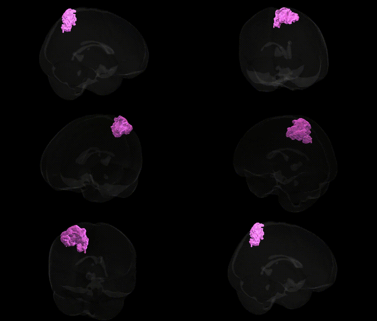
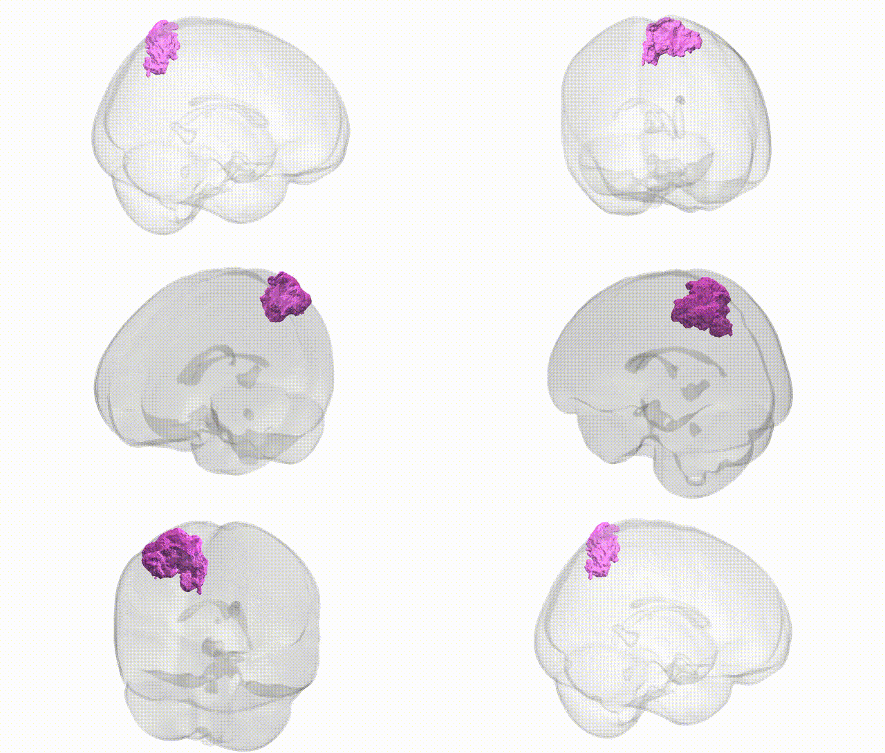
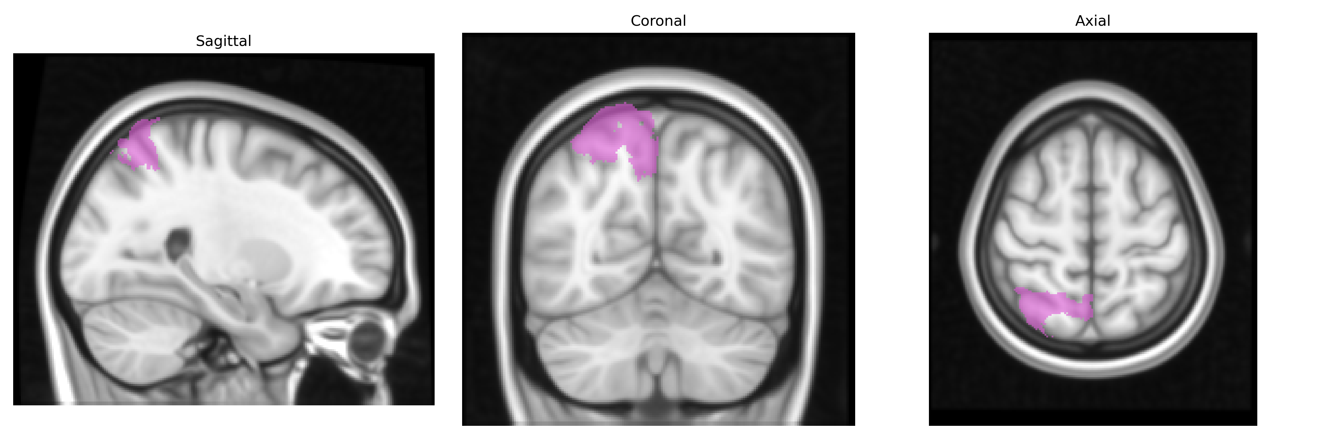
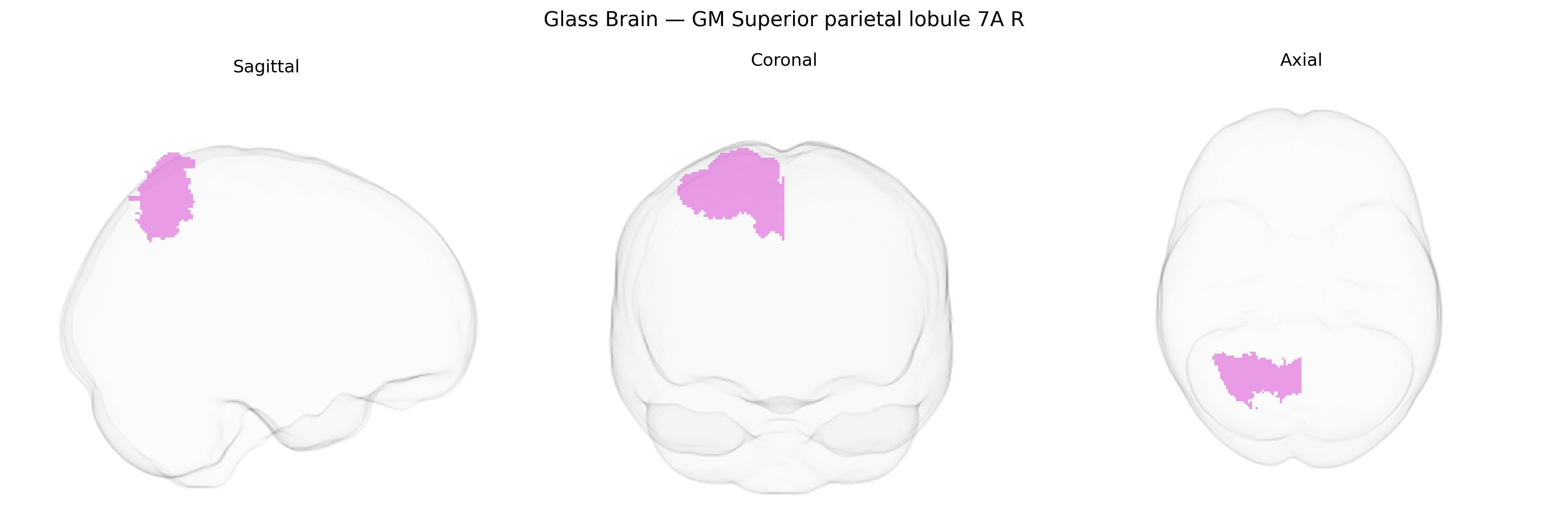
The bilateral GM Superior parietal lobule 7A R, as defined in the Juelich maxprob thr25 1mm atlas, corresponds to a cytoarchitectonically defined subregion of the superior parietal lobule located in the posterior parietal cortex, dorsally and medially on the lateral surface of the hemisphere. This area is implicated in higher-order sensorimotor integration, visuospatial processing, and the coordination of attention and eye–hand movements, integrating visual, somatosensory, and proprioceptive inputs for the guidance of actions in space. Functionally, it contributes to spatial orientation, mental rotation, and the internal representation of body position in relation to the external environment, supporting complex goal-directed behavior and aspects of working memory for spatial information. There is no direct Wikipedia article for “Area 7A (Juelich)”; a related structure is the Superior parietal lobule.
The bilateral GM Superior parietal lobule 7A (right hemisphere) in the Juelich maxprob thr25 1mm atlas corresponds to a higher-order parietal association area implicated in visuospatial attention, sensorimotor integration, and aspects of working memory, and genetic associations to this region largely arise from imaging-genetics and GWAS of brain structure and function rather than region-specific candidate-gene work. Large neuroimaging GWAS consortia (e.g., ENIGMA, UK Biobank) have reported heritable variation in parietal cortical thickness, surface area, and gyrification, with common variants near genes involved in neurodevelopment, synaptic function, and cytoskeletal organization (such as MAPT, DLG2, and genes in Wnt and axon-guidance pathways) contributing to individual differences in parietal morphology, some of which overlap with general cognitive ability and educational attainment loci. Functional neuroimaging GWAS has linked genetic variation in glutamatergic, dopaminergic, and GABAergic signaling genes to activation patterns in superior parietal regions during attention and working-memory tasks, while polygenic scores for intelligence and cognitive performance show associations with superior parietal volume and activity. Clinically, parietal lobule structure and connectivity—particularly in superior parietal areas—have been associated in GWAS and polygenic-risk studies with traits and disorders such as ADHD, schizophrenia, major depressive disorder, and autism spectrum conditions, often as part of distributed frontoparietal and default-mode networks rather than isolated 7A-specific effects. Specific Juelich-defined area 7A R has not yet been the direct focus of large, high-resolution GWAS, and most genetic associations are inferred from broader parietal or superior parietal measures, but converging evidence supports a role of common genetic variation influencing its morphology and function in relation to attentional control, visuospatial processing, and general cognitive ability, with overlapping polygenic architectures across neurodevelopmental and psychiatric conditions.
Overview generated by GPT-4o (2026).
Region ID: 74
Hemisphere: bilateral
Atlas: Juelich maxprob thr25 1mm
Full Quality Version: Download MP4
Full Quality Version: Download MP4
Wali Sidiqyar*, Gaurav Rudravaram*, Elyssa M. McMaster, Trent M. Schwartz, Adam M. Saunders, Kurt G. Schilling, Bennett A. Landman "Introducing SPINS: A Shared Public Visualization Library of Neuroanatomical Structures." Medical Imaging with Deep Learning- short paper
This resource is licensed under CC0 1.0 Universal (Public Domain).