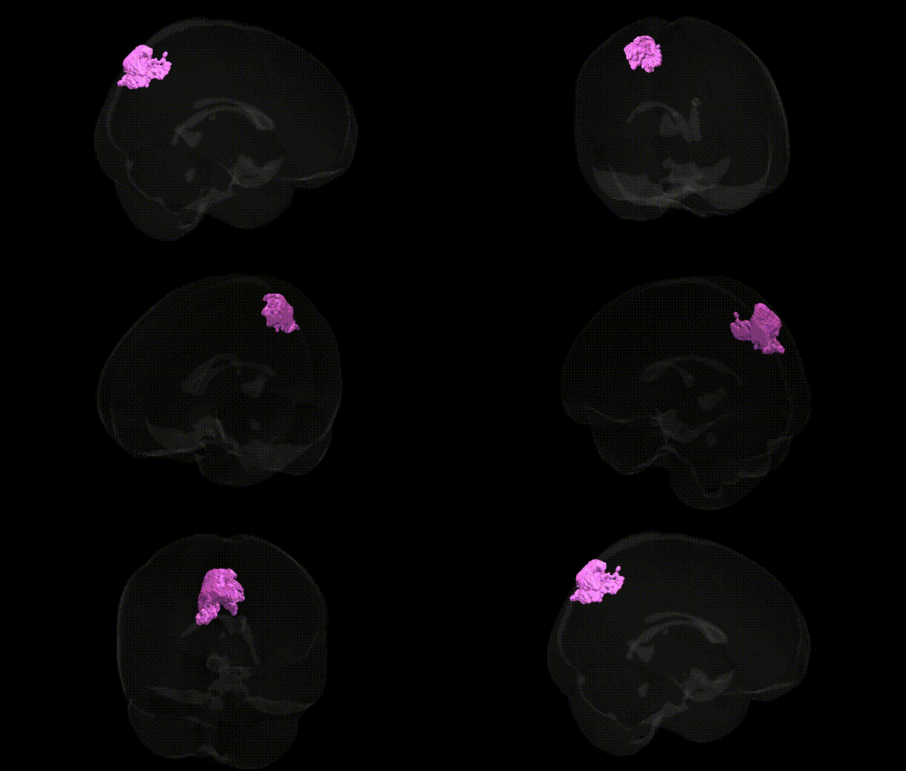
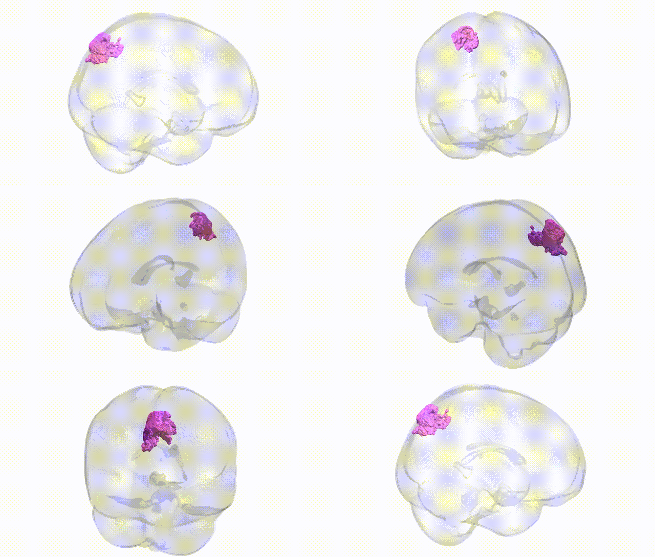
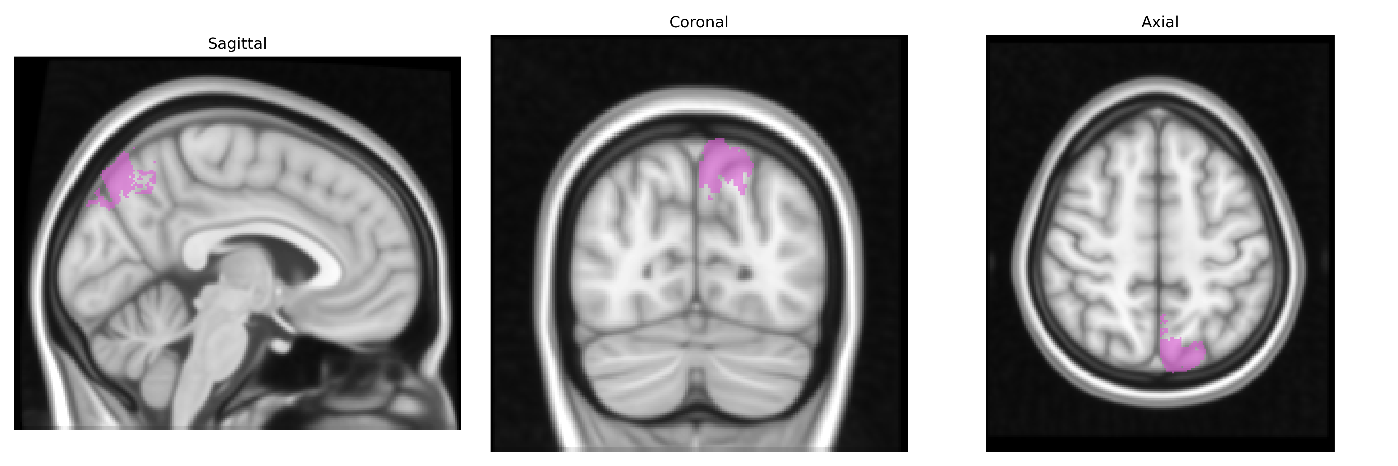
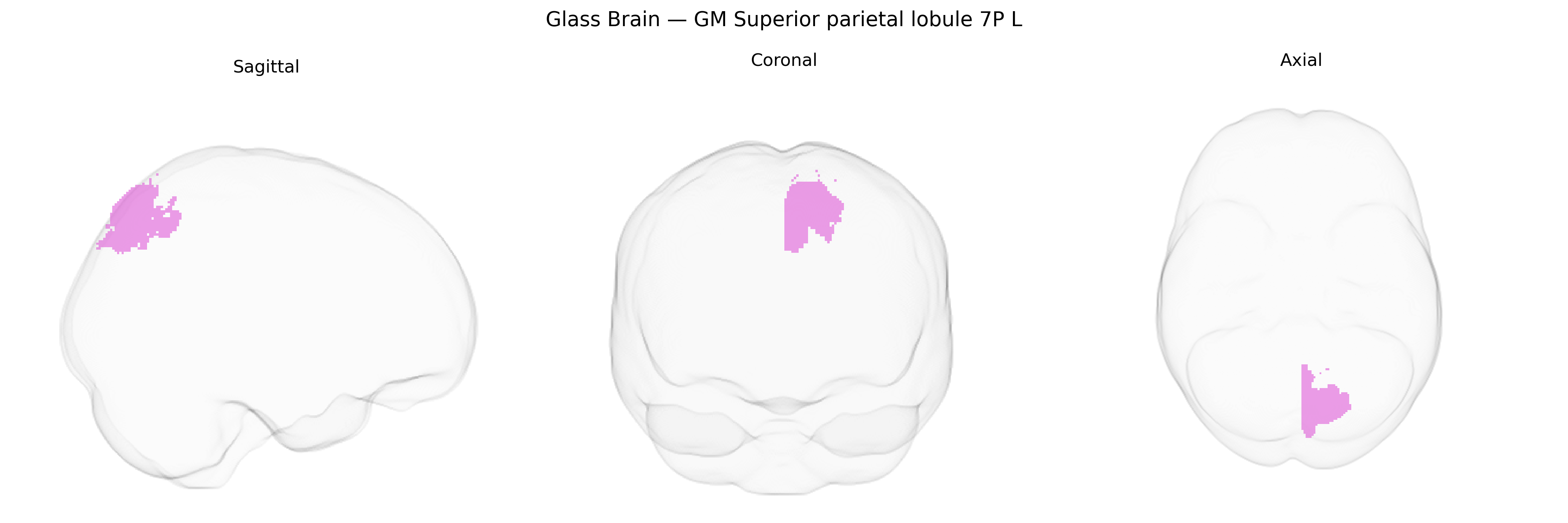
The bilateral GM Superior parietal lobule 7P L, as defined in the Juelich maxprob thr25 1mm atlas, corresponds to a cytoarchitectonic subdivision of Brodmann area 7 located in the superior parietal lobule of the left hemisphere. This region is implicated in higher-order somatosensory processing, visuospatial integration, and sensorimotor transformations supporting functions such as spatial attention, body-centered coordinate mapping, and visually guided reaching and grasping. Neuronal populations in 7P integrate multimodal inputs from visual, somatosensory, and premotor areas, contributing to the construction and maintenance of spatial representations and the coordination of goal-directed actions. There is no direct link for area 7P; a related structure is the superior parietal lobule: Superior parietal lobule.
The bilateral grey matter Superior Parietal Lobule area 7P (left hemisphere) from the Juelich maxprob atlas is part of a parietal association network implicated in visuospatial integration, attention, and higher-order sensorimotor processing, and genetic associations involving this region largely emerge from imaging-genetics and GWAS of cortical structure and functional connectivity rather than from studies targeting 7P specifically. Large neuroimaging GWAS consortia (e.g., ENIGMA, UK Biobank) have identified multiple common variants in genes related to neuronal development and synaptic function (such as variants near HMGA2, KIAA0586, THBS3, and IGF1) that are associated with parietal cortical thickness, surface area, and gyrification, measures that include or overlap superior parietal territories. Polygenic risk scores for neurodevelopmental and psychiatric conditions—including schizophrenia, ADHD, and autism spectrum disorder—have been associated with altered parietal morphology and connectivity, with superior parietal regions frequently showing differences in case–control and risk-carrier samples, though these are generally network-level rather than 7P-specific findings. GWAS of cognitive traits such as general intelligence, working memory, and educational attainment have implicated genetic loci that also correlate with structural and functional variation in parietal association cortex, supporting a genetically influenced role of this region in higher cognition. Additionally, variants linked to Alzheimer’s disease and other dementias (e.g., in APOE and CLU) show associations with atrophy and hypometabolism in parietal association areas, including the superior parietal lobule, in imaging-genetics studies. Overall, current evidence indicates that genetic influences on neurodevelopment, synaptic organization, and neurodegeneration shape structure and function of the superior parietal lobule, but specific GWAS focused narrowly on Juelich’s 7P subregion are not yet available, and most genetic findings relate to broader parietal or frontoparietal networks.
Overview generated by GPT-4o (2026).
Region ID: 79
Hemisphere: bilateral
Atlas: Juelich maxprob thr25 1mm
Full Quality Version: Download MP4
Full Quality Version: Download MP4
Wali Sidiqyar*, Gaurav Rudravaram*, Elyssa M. McMaster, Trent M. Schwartz, Adam M. Saunders, Kurt G. Schilling, Bennett A. Landman "Introducing SPINS: A Shared Public Visualization Library of Neuroanatomical Structures." Medical Imaging with Deep Learning- short paper
This resource is licensed under CC0 1.0 Universal (Public Domain).