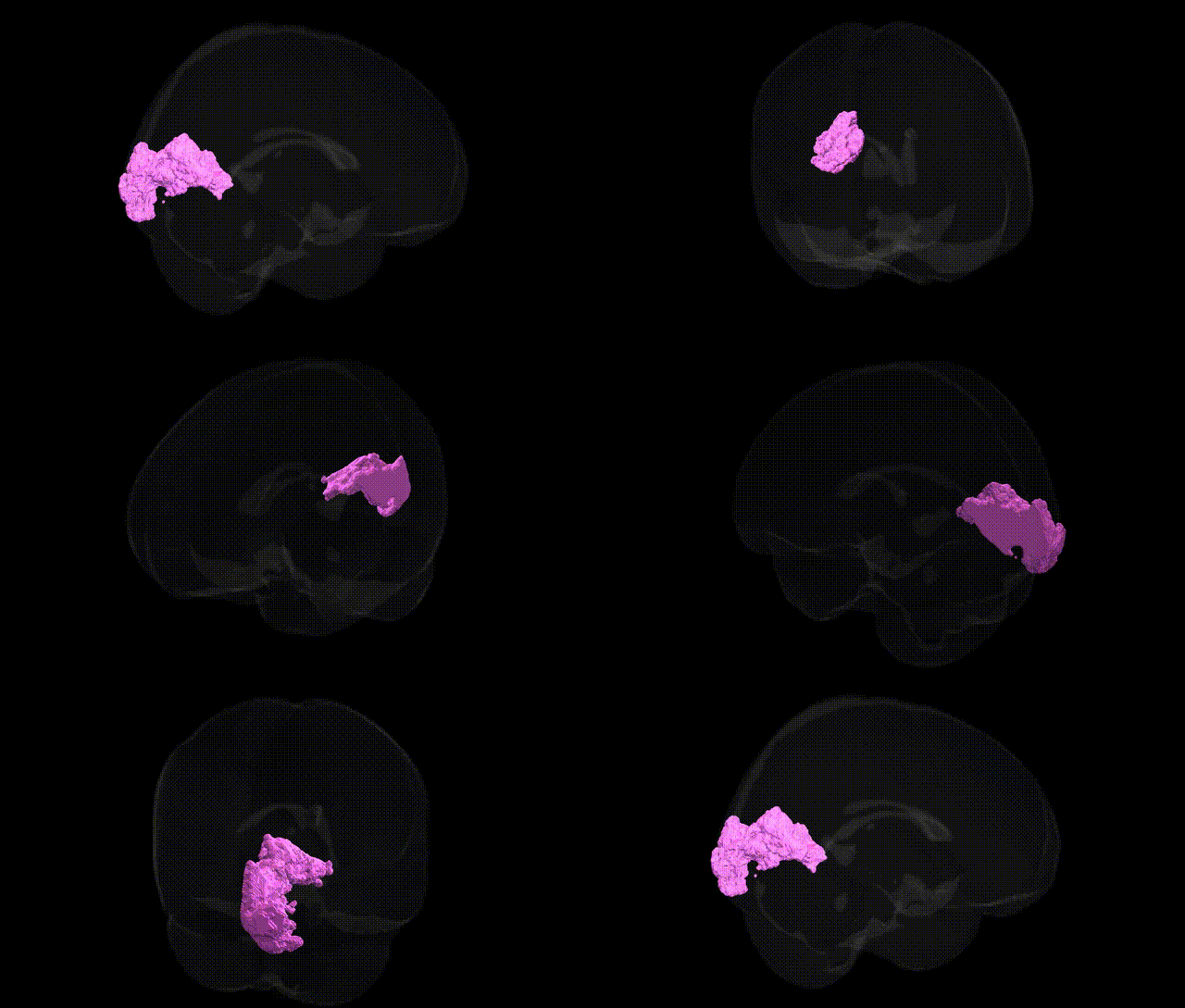
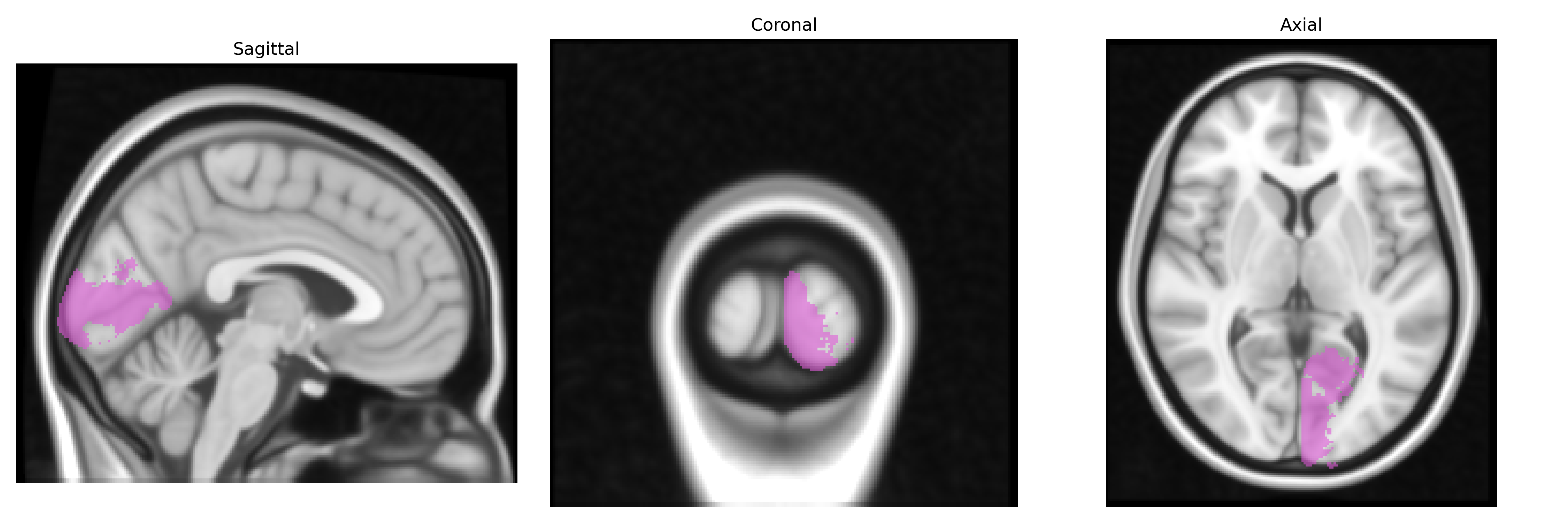
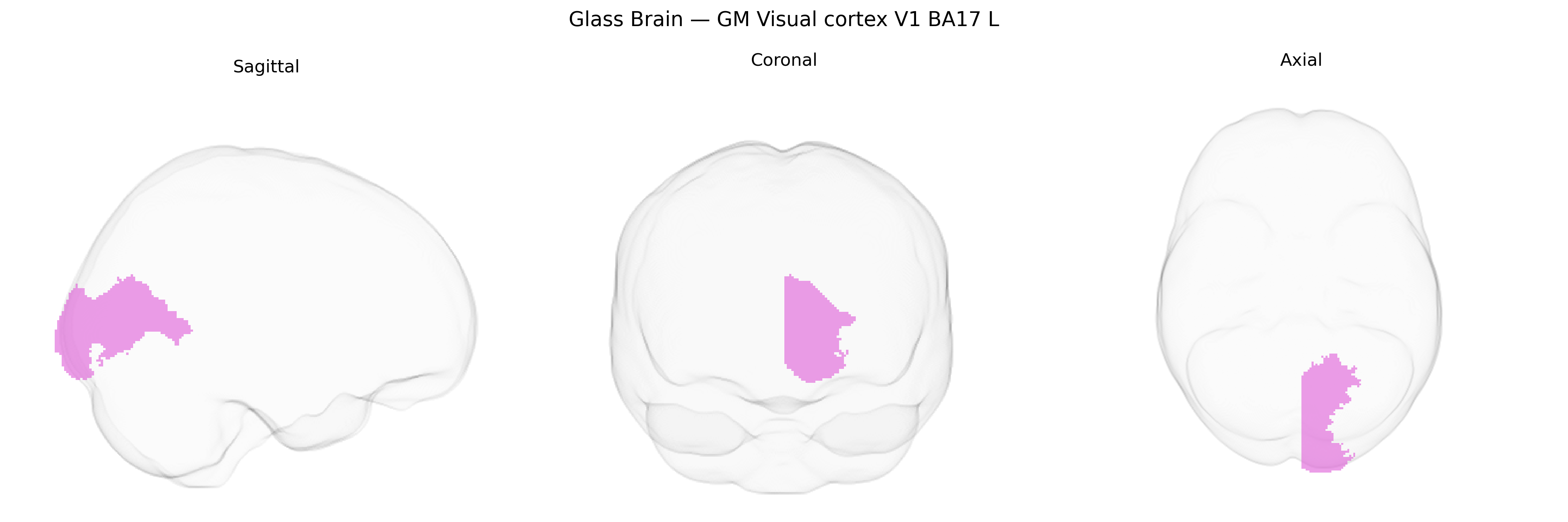
The bilateral GM Visual cortex V1 BA17 L region from the Juelich maxprob thr25 1mm atlas corresponds to the left primary visual cortex, situated predominantly along the banks of the calcarine sulcus in the occipital lobe and cytoarchitectonically defined as Brodmann area 17. This region contains a dense granular layer IV, receives topographically organized input from the lateral geniculate nucleus of the thalamus, and is characterized by a retinotopic map in which central vision is represented posteriorly and peripheral vision more anteriorly. Neurons within V1 show orientation, ocular dominance, and spatial frequency selectivity, forming columnar and laminar microcircuits that support the initial cortical processing of visual information, including edge detection, contrast analysis, and basic feature extraction, which are subsequently relayed to extrastriate visual areas for higher-order processing. There is no direct Wikipedia article for the specific Juelich-defined “Visual cortex V1 BA17 L” region; a closely related entry describing the same functional and anatomical territory is Visual cortex.
Genetic associations involving the bilateral primary visual cortex (V1, Brodmann area 17, as defined in the Juelich maxprob thr25 1mm atlas) arise largely from imaging–genetics and GWAS studies of cortical thickness, surface area, and functional activation rather than region-specific candidate-gene work; polygenic influences linked to general brain morphometry, such as variants in genes regulating neurodevelopment, synaptic plasticity, and myelination (including loci near genes like HMGA2, MIR588, and others identified in large ENIGMA and UK Biobank analyses), contribute to V1 structural variance. GWAS of visual cortex measures have connected this region to global cognitive and educational traits, reflecting pleiotropy between occipital morphology and intelligence or educational attainment, and to cross-trait genetic overlap with psychiatric and neurodevelopmental disorders (e.g., schizophrenia, major depression, autism spectrum conditions), in which altered V1 thickness or surface area is frequently reported. Additional associations emerge in studies of albinism and other congenital visual pathway disorders where genes affecting retinal and optic nerve development (e.g., TYR, OCA2, GPR143) indirectly alter V1 structure and organization, as well as in migraine and visual aura research implicating occipital excitability, though specific common-risk variants mapping exclusively or predominantly to BA17 remain limited and typically form part of broader polygenic architectures influencing multiple cortical regions.
Overview generated by GPT-4o (2026).
Region ID: 81
Hemisphere: bilateral
Atlas: Juelich maxprob thr25 1mm
Full Quality Version: Download MP4
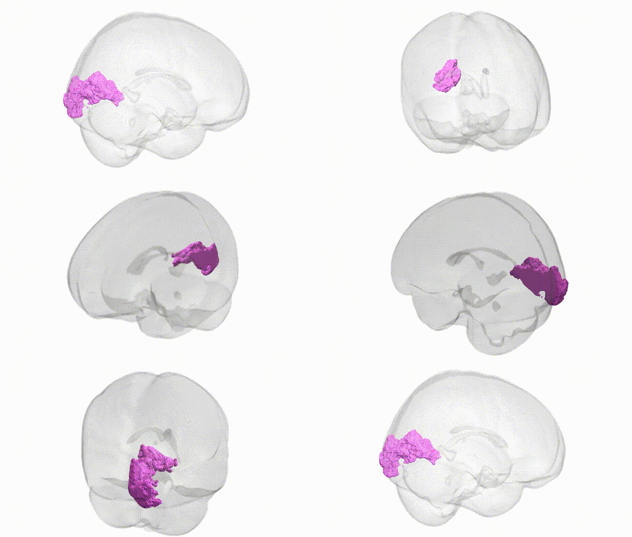
Full Quality Version: Download MP4
Wali Sidiqyar*, Gaurav Rudravaram*, Elyssa M. McMaster, Trent M. Schwartz, Adam M. Saunders, Kurt G. Schilling, Bennett A. Landman "Introducing SPINS: A Shared Public Visualization Library of Neuroanatomical Structures." Medical Imaging with Deep Learning- short paper
This resource is licensed under CC0 1.0 Universal (Public Domain).