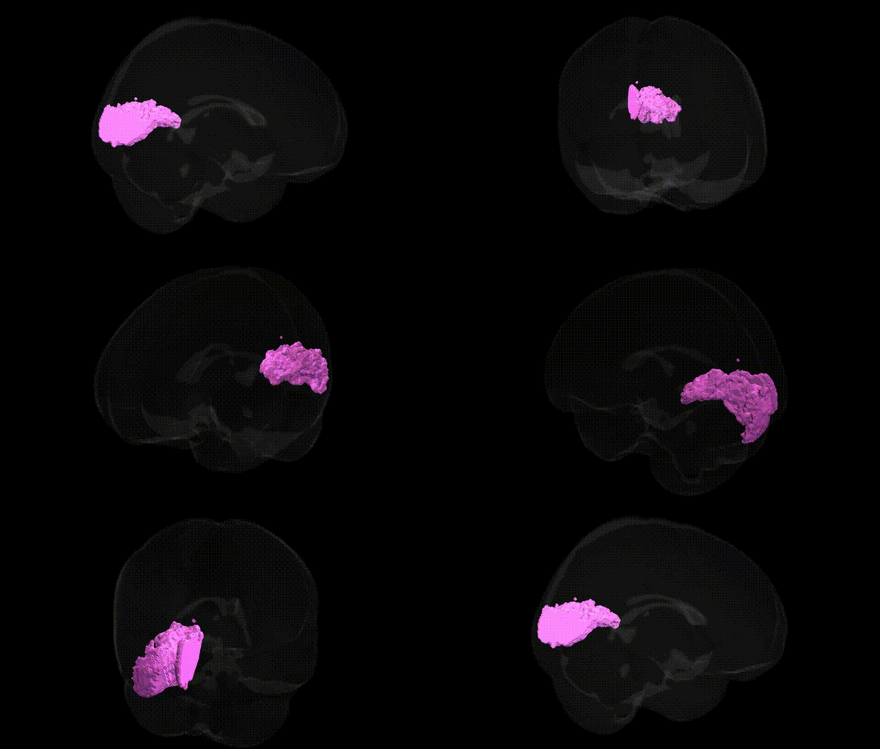
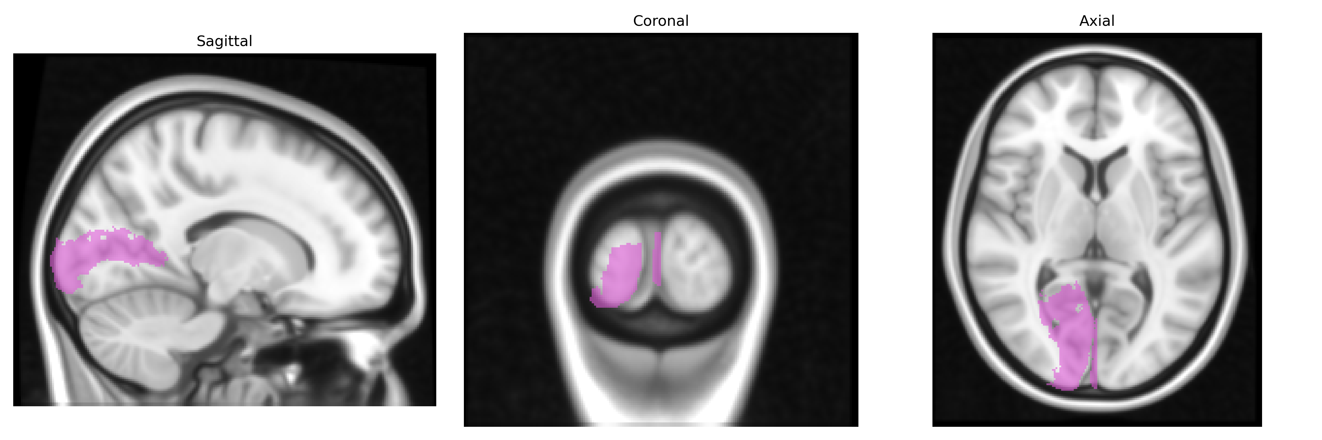
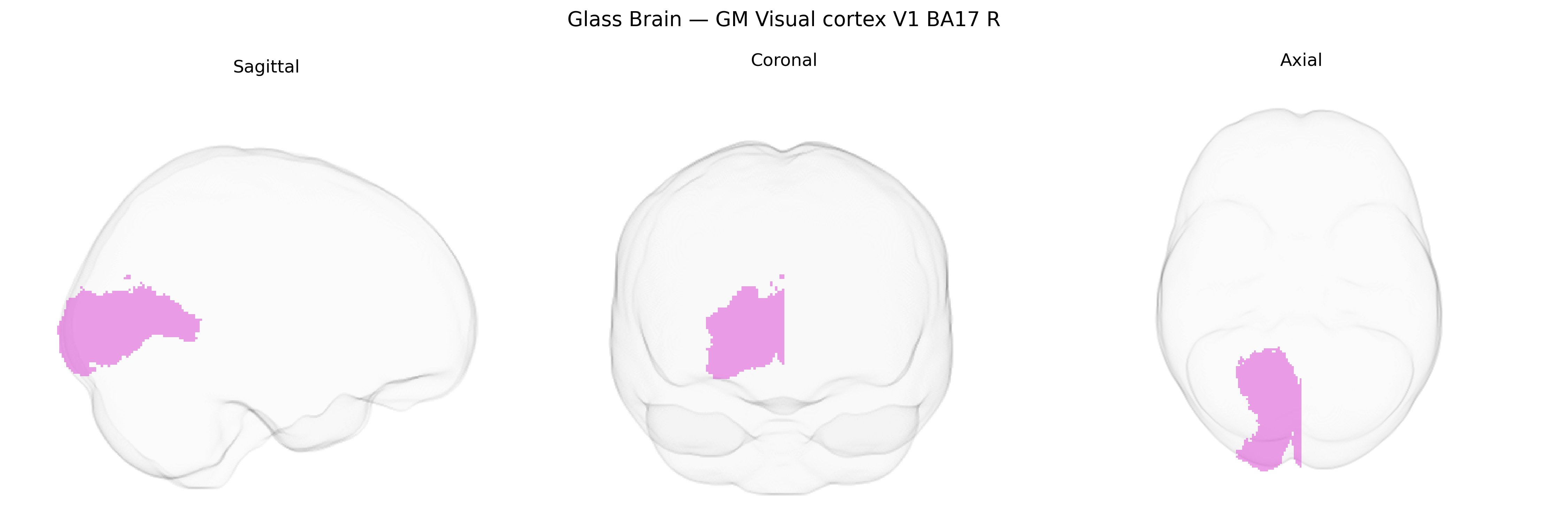
The bilateral GM Visual cortex V1 BA17 R region from the Juelich maxprob thr25 1mm Atlas corresponds to the right primary visual cortex, a cytoarchitectonically defined area located along the calcarine sulcus in the occipital lobe and classically identified as Brodmann area 17. This region receives the densest projections from the lateral geniculate nucleus of the thalamus and serves as the main cortical entry point for visual information, exhibiting a precise retinotopic organization in which neighboring neurons represent neighboring points in the visual field, with a disproportionate cortical representation of the fovea. Neurons in this region are highly specialized for basic visual features such as orientation, spatial frequency, contrast, and motion direction, forming columns and layers that support early-stage visual processing and feedforward projections to extrastriate visual areas. Functionally, the right-hemisphere V1 is critical for processing visual input from the left visual field and for generating the initial cortical representation of visual scenes that underlies higher-order perception and visually guided behavior. Primary visual cortex
The bilateral GM visual cortex V1 (BA17, right hemisphere) in the Juelich maxprob atlas has been implicated in several genetic association frameworks, mainly through imaging genetics and large-scale GWAS of brain structure and function. Variants in genes involved in synaptic plasticity, neuronal development, and visual processing—such as BDNF, GRIN2B, NRG1, and genes influencing glutamatergic and GABAergic signaling—have been associated with individual differences in V1 cortical thickness, surface area, and activity in response to visual stimuli. ENIGMA and UK Biobank–based GWAS of cortical morphology have identified multiple common variants with small effect sizes that influence occipital and primary visual cortex anatomy, some overlapping with loci implicated in general cognitive ability and educational attainment. Genetic risk for neurodevelopmental and psychiatric disorders including schizophrenia, autism spectrum disorder, and major depression has been linked to altered V1 structure or function, with polygenic risk scores for these conditions showing associations with occipital cortical metrics. In addition, rare variants and copy-number changes impacting genes involved in early cortical patterning (e.g., transcription factors and axon guidance molecules) have been reported in disorders featuring visual perceptual anomalies or visual field deficits, indirectly implicating V1. Overall, genetic influences on V1 are highly polygenic and pleiotropic, with many loci contributing modestly to variability in its structure and visual processing and overlapping substantially with broader neuropsychiatric and cognitive trait genetics.
Overview generated by GPT-4o (2026).
Region ID: 82
Hemisphere: bilateral
Atlas: Juelich maxprob thr25 1mm
Full Quality Version: Download MP4
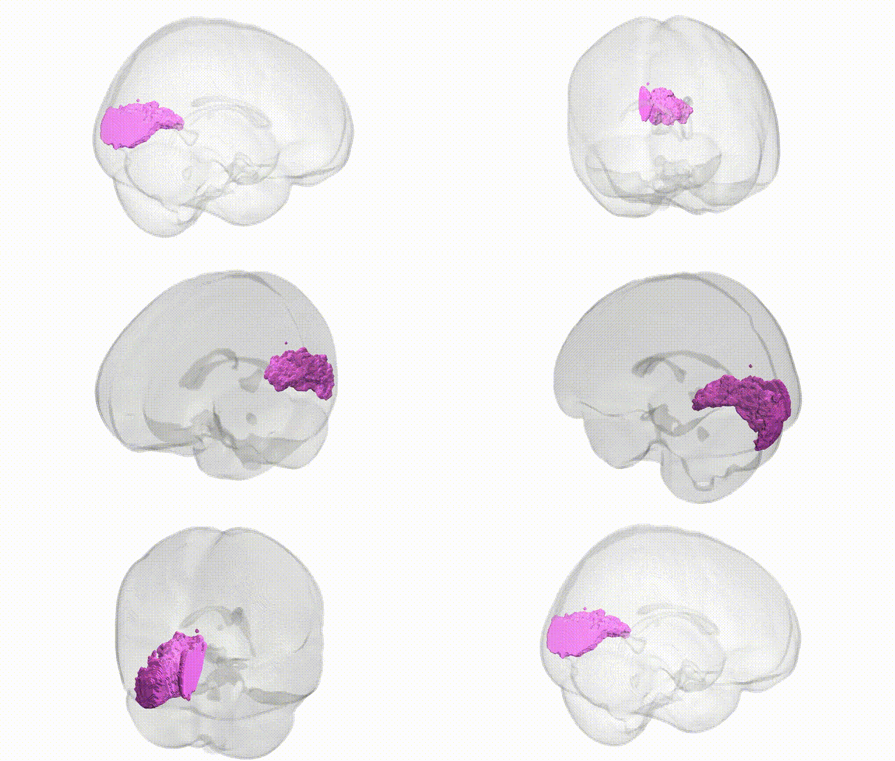
Full Quality Version: Download MP4
Wali Sidiqyar*, Gaurav Rudravaram*, Elyssa M. McMaster, Trent M. Schwartz, Adam M. Saunders, Kurt G. Schilling, Bennett A. Landman "Introducing SPINS: A Shared Public Visualization Library of Neuroanatomical Structures." Medical Imaging with Deep Learning- short paper
This resource is licensed under CC0 1.0 Universal (Public Domain).