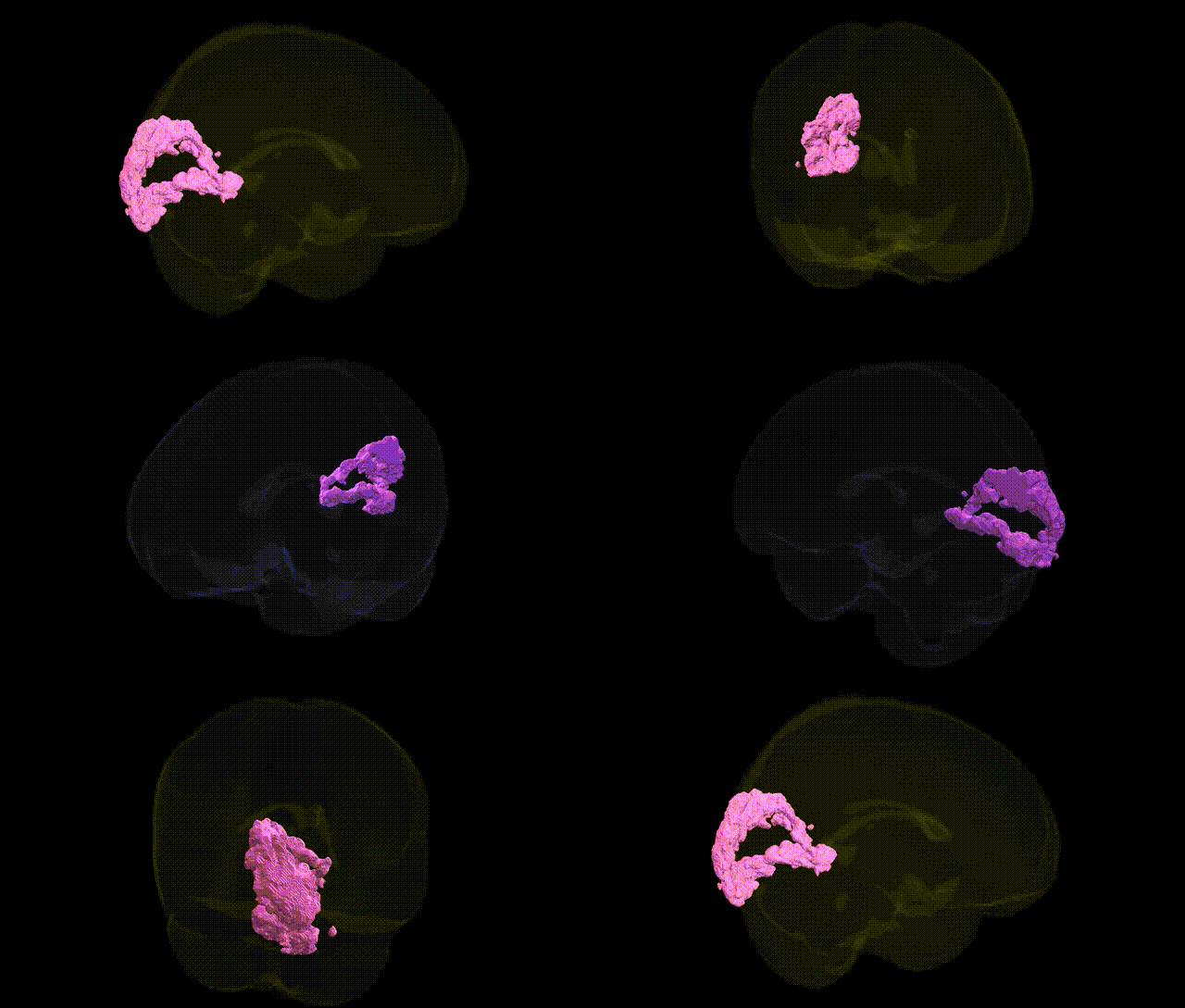
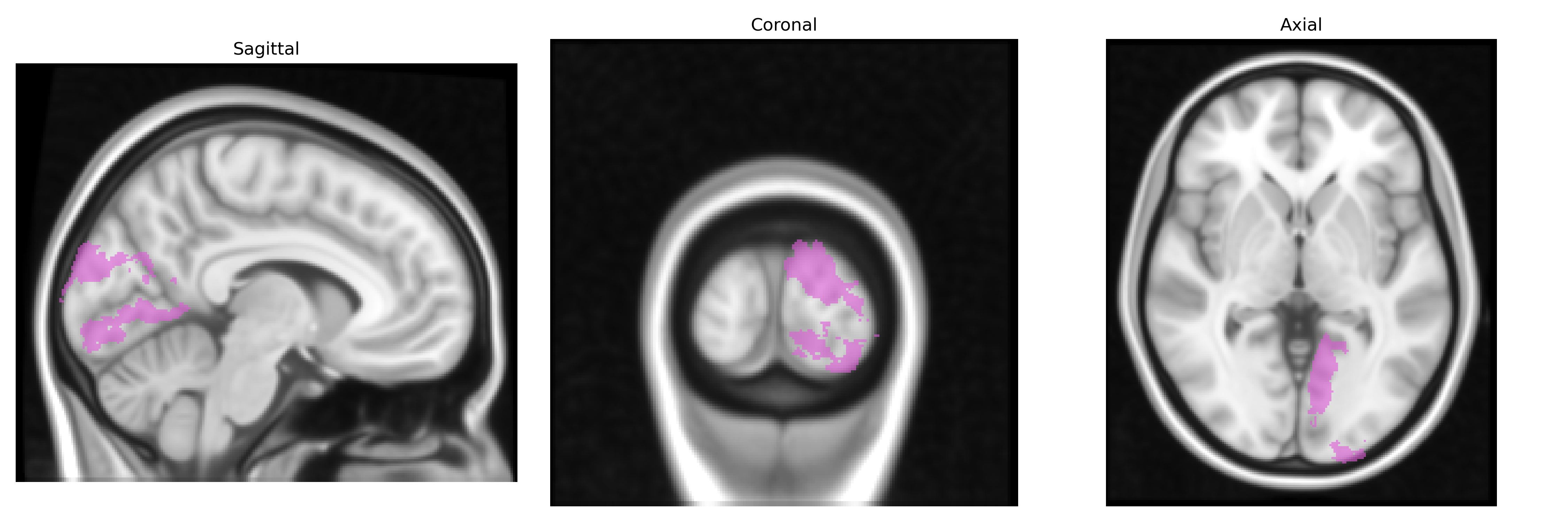
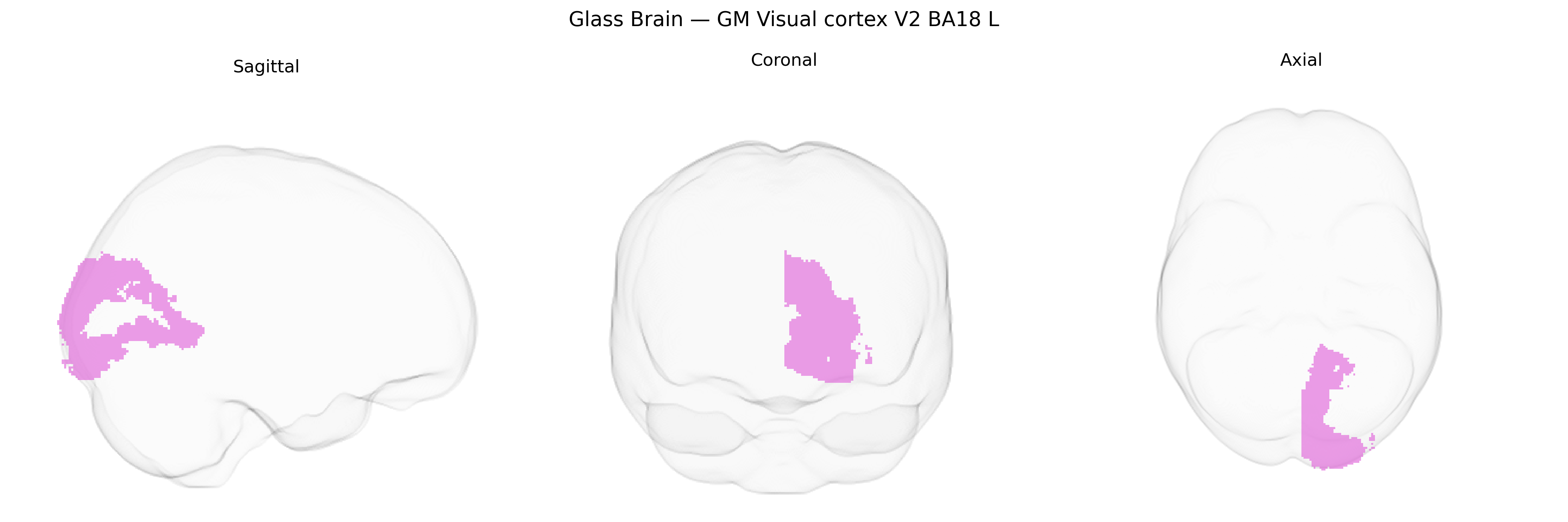
The bilateral GM Visual cortex V2 BA18 L region from the Juelich maxprob thr25 1mm Atlas corresponds to the left-hemispheric portion of Brodmann area 18, a secondary visual area (V2) located in the occipital lobe adjacent to primary visual cortex (V1/BA17). This region is part of early extrastriate visual cortex and receives strong feedforward input from V1, as well as feedback and lateral connections from higher-order visual areas. Neurons in BA18/V2 are involved in processing more complex visual features than V1, including orientation combinations, contours, figure–ground segmentation, binocular disparity, and basic motion and color patterns, contributing to both dorsal (“where/how”) and ventral (“what”) visual processing streams. Functionally, this area participates in early stages of object recognition, spatial organization, and integration of visual information across the visual field, with retinotopic organization preserved from V1 but with larger receptive fields and increasing invariance. There is no direct Wikipedia article for this exact atlas-defined region; a closely related structure is Brodmann area 18.
The bilateral grey matter visual cortex V2 (BA18, left hemisphere) from the Juelich maxprob thr25 1mm atlas participates in intermediate visual processing and has been implicated in several genetic and neuroimaging–genetic findings, although few studies target BA18 specifically. Large-scale GWAS of cortical structure (e.g., ENIGMA and UK Biobank) have identified common variants in multiple genes, including those related to synaptic function and neurodevelopment (such as MAPT, POU3F2, and genes in glutamatergic and GABAergic pathways), that influence occipital cortical thickness and surface area encompassing BA18/V2. Polygenic risk scores for schizophrenia, bipolar disorder, and major depression have been associated with structural and functional changes in occipital visual regions, including V2, while autism spectrum disorder and dyslexia studies have linked genetic variation in neuronal migration and axon guidance genes (e.g., CNTNAP2, DCDC2) to altered visual cortex activation patterns during perceptual and reading tasks. GWAS of visual perception and cognitive traits (such as general intelligence, reading ability, and face recognition) have identified variants near genes involved in cortical development (e.g., HMGA2, KIAA0319, FOXP2-related networks) that show downstream associations with activity and morphology in early visual cortices, including BA18. In addition, migraine and visual aura genetic studies have reported associations in ion channel and neurotransmission genes (CACNA1A and others) that are linked to excitability differences in occipital visual cortex, while GWAS of resting-state functional connectivity have connected variants in neurodevelopmental and synaptic genes to altered connectivity between BA18/V2 and higher-order visual and parietal networks. Overall, genetic influences on BA18/V2 appear to be distributed across many loci affecting cortical development, synaptic signaling, and excitability, with convergent evidence from GWAS and imaging-genetics linking this region to psychiatric risk, neurodevelopmental traits, and visual and cognitive performance.
Overview generated by GPT-4o (2026).
Region ID: 83
Hemisphere: bilateral
Atlas: Juelich maxprob thr25 1mm
Full Quality Version: Download MP4
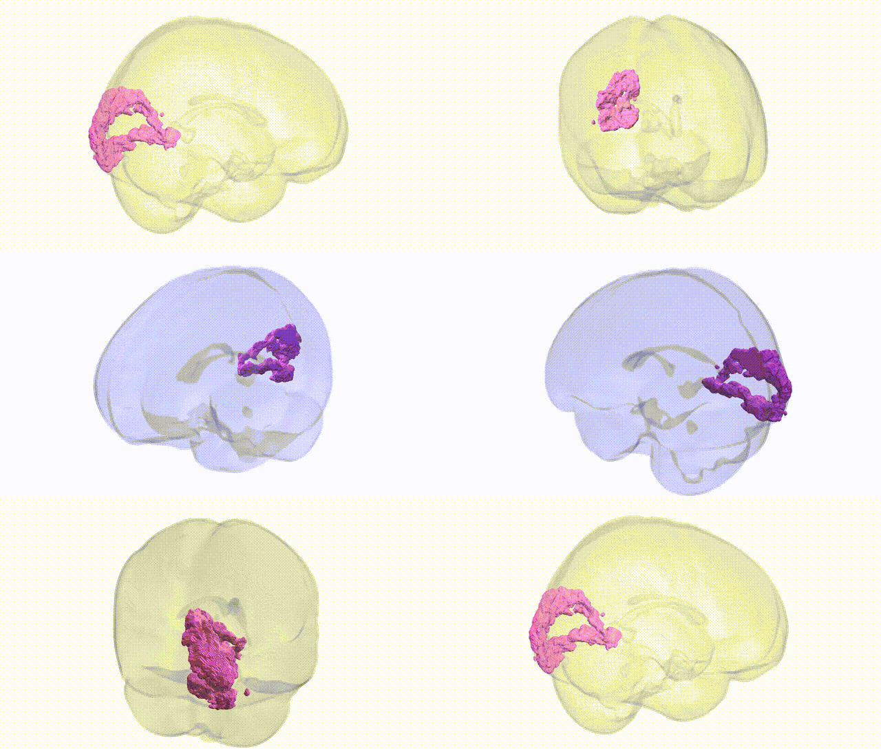
Full Quality Version: Download MP4
Wali Sidiqyar*, Gaurav Rudravaram*, Elyssa M. McMaster, Trent M. Schwartz, Adam M. Saunders, Kurt G. Schilling, Bennett A. Landman "Introducing SPINS: A Shared Public Visualization Library of Neuroanatomical Structures." Medical Imaging with Deep Learning- short paper
This resource is licensed under CC0 1.0 Universal (Public Domain).