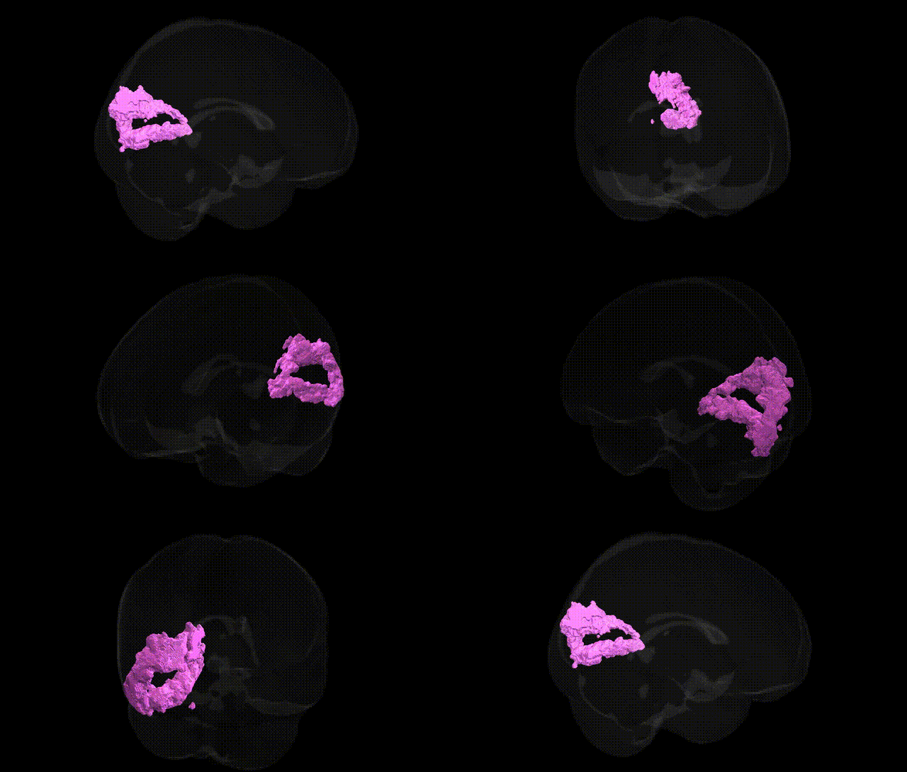
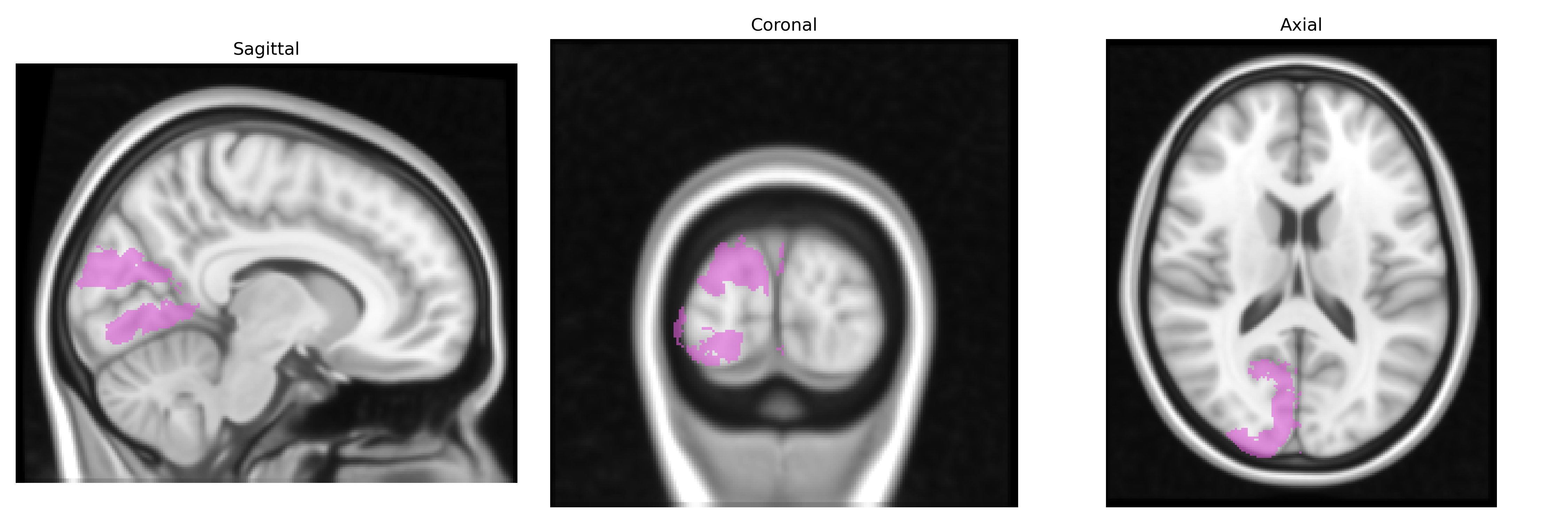
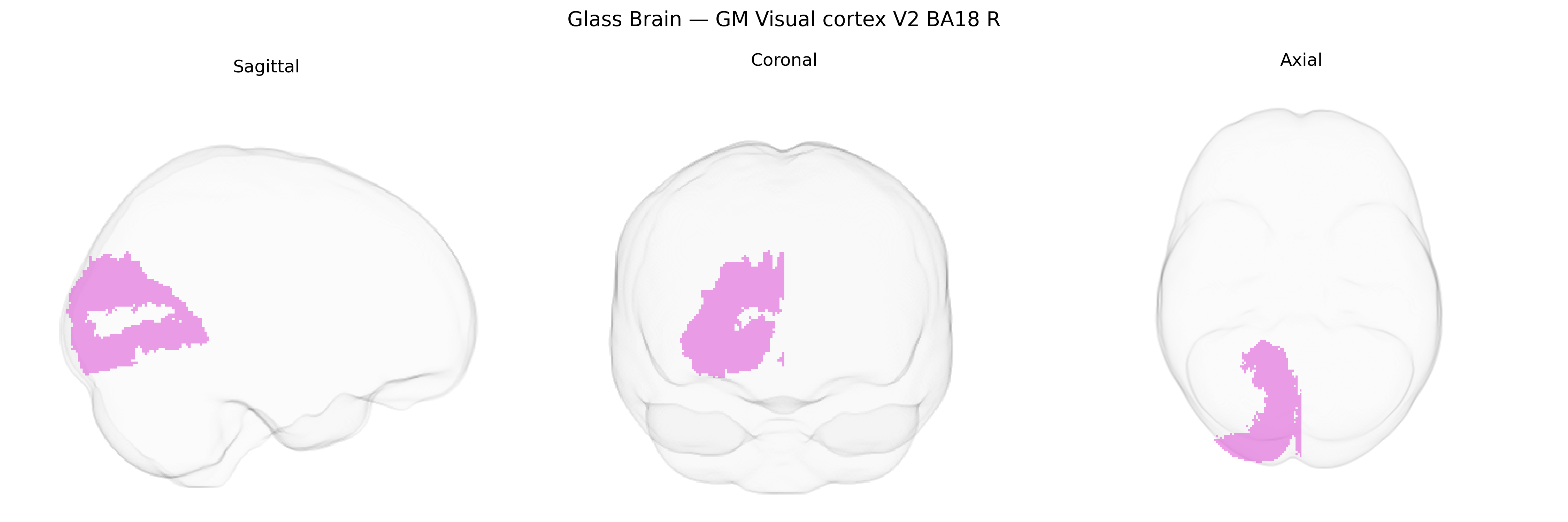
The bilateral GM Visual cortex V2 BA18 R region from the Juelich maxprob thr25 1 mm atlas corresponds to cytoarchitectonic area 18 in the right (and symmetrically left) occipital lobe, representing the secondary visual cortex (V2) situated adjacent to primary visual cortex (V1, BA17) along the calcarine fissure. This region forms an early-stage visual processing hub that receives strong feedforward input from V1 and projects to higher-order extrastriate areas, participating in the analysis of orientation, spatial frequency, binocular disparity, figure–ground segregation, and contour integration. Neuronal populations in BA18/V2 exhibit columnar and stripe-like organization (thin, thick, and pale stripes) that support parallel processing of color, motion, and form information, providing critical integration for dorsal and ventral visual streams. Functionally, this area contributes to visual perception, pattern recognition, and the transformation of low-level retinal input into more complex representations necessary for object, scene, and motion processing. Visual area V2
The bilateral GM visual cortex V2 (BA18, right hemisphere) in the Juelich maxprob atlas has been implicated in genetic studies primarily through large-scale neuroimaging GWAS that link common variants to regional cortical thickness, surface area, and functional activation in visual processing tasks. Variants near genes involved in synaptic development and visual pathway function (including but not limited to GABRB3, NRXN1, GRIN2B, and genes in calcium signaling and neurodevelopmental pathways) have been associated with structural and functional measures in occipital visual areas encompassing V2/BA18. GWAS of brain morphology from consortia such as ENIGMA and UK Biobank report heritable variation in occipital cortical metrics, with polygenic influences overlapping those for cognitive performance, educational attainment, and general brain size. V2/BA18-related occipital regions show genetic correlations with neurodevelopmental and psychiatric conditions that feature visual-perceptual or attentional abnormalities, including schizophrenia, autism spectrum disorder, ADHD, and migraine with aura, often via pleiotropic loci affecting excitatory–inhibitory balance and visual network connectivity. Functional genetic imaging studies have identified associations between common polymorphisms in dopaminergic and serotonergic genes (e.g., COMT, 5-HTTLPR) and modulation of visual cortex responses, while rare variant and CNV studies highlight broad occipital involvement in syndromic disorders with visual processing deficits rather than specific V2-localized effects. Overall, genetic findings support a heritable architecture influencing V2/BA18 structure and function, embedded within wider occipital and multimodal networks that contribute to perception, cognition, and risk for neuropsychiatric disorders.
Overview generated by GPT-4o (2026).
Region ID: 84
Hemisphere: bilateral
Atlas: Juelich maxprob thr25 1mm
Full Quality Version: Download MP4
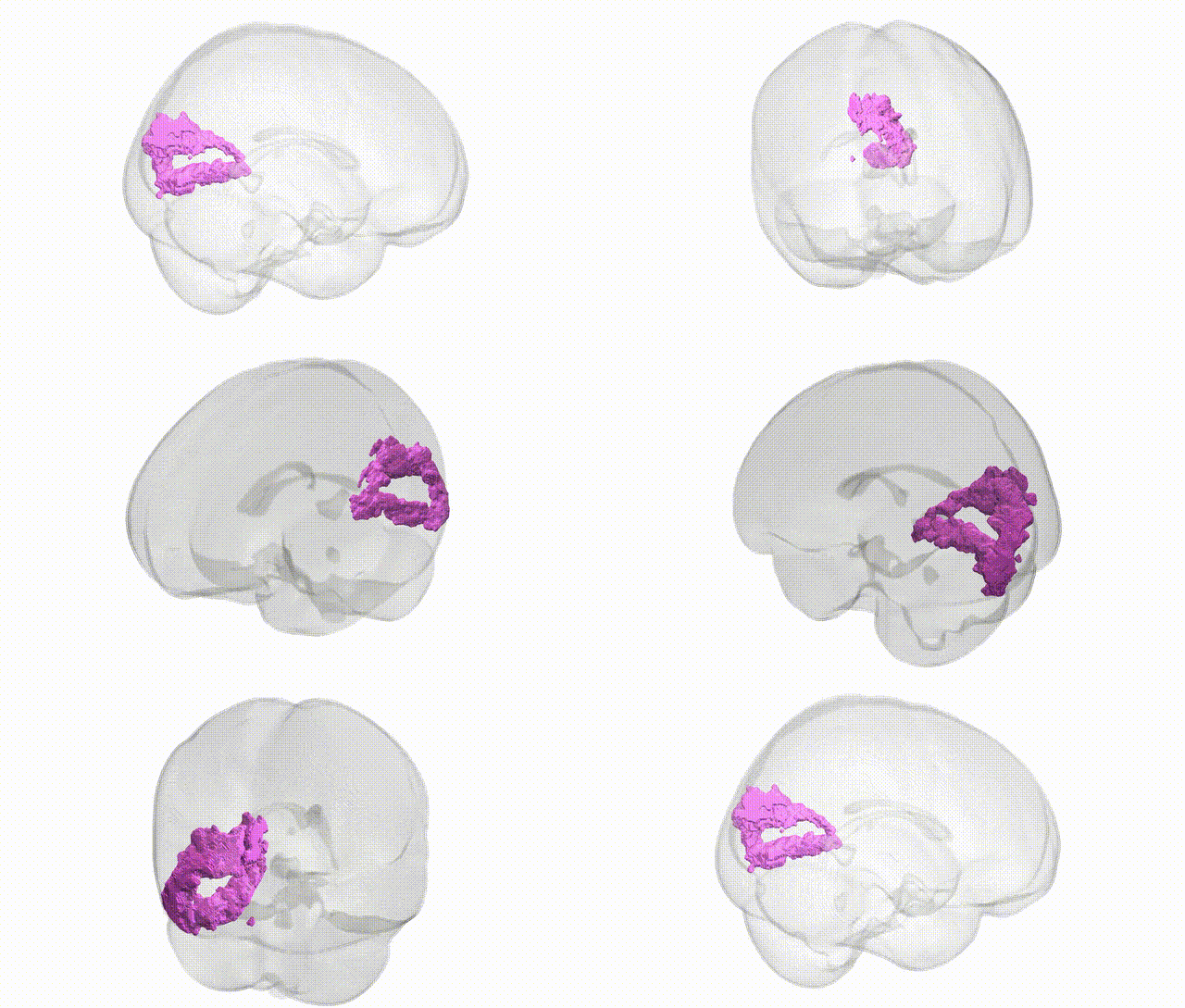
Full Quality Version: Download MP4
Wali Sidiqyar*, Gaurav Rudravaram*, Elyssa M. McMaster, Trent M. Schwartz, Adam M. Saunders, Kurt G. Schilling, Bennett A. Landman "Introducing SPINS: A Shared Public Visualization Library of Neuroanatomical Structures." Medical Imaging with Deep Learning- short paper
This resource is licensed under CC0 1.0 Universal (Public Domain).