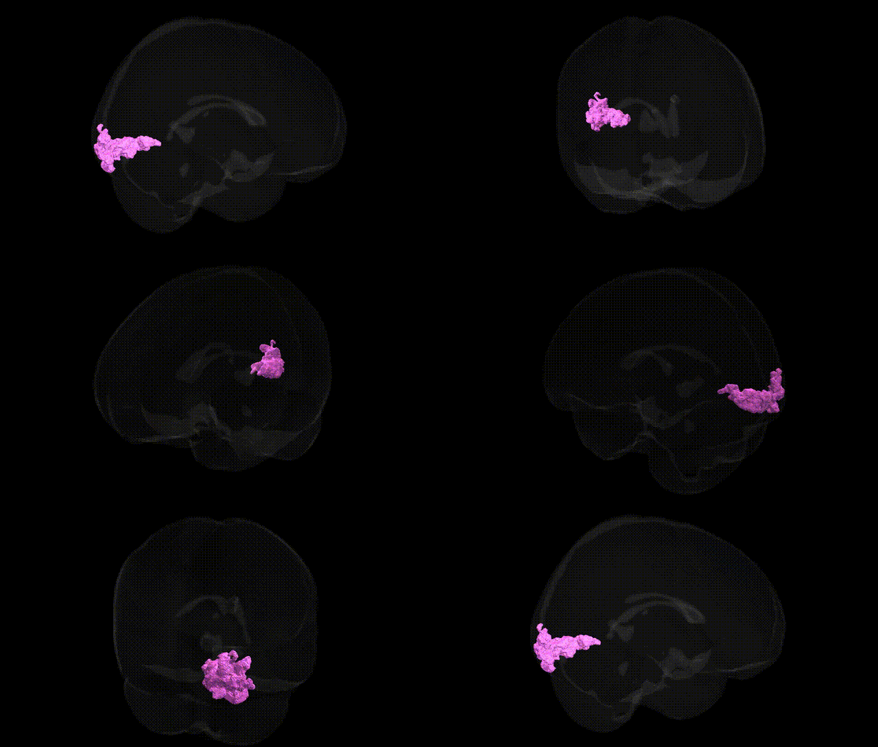
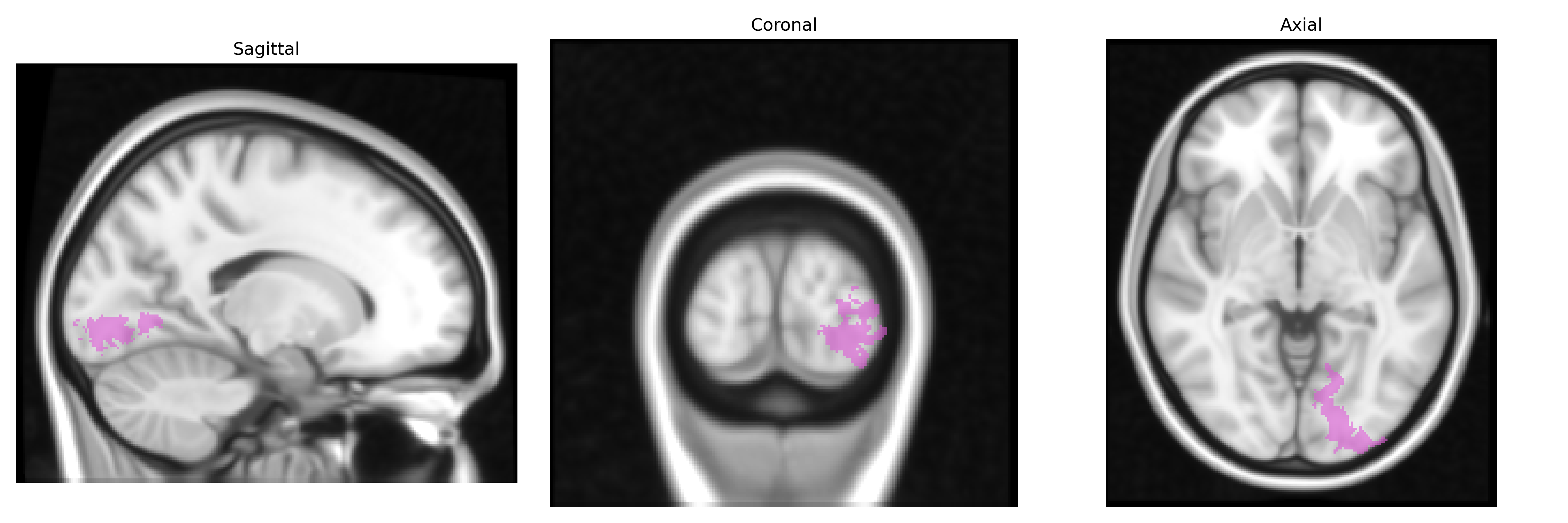
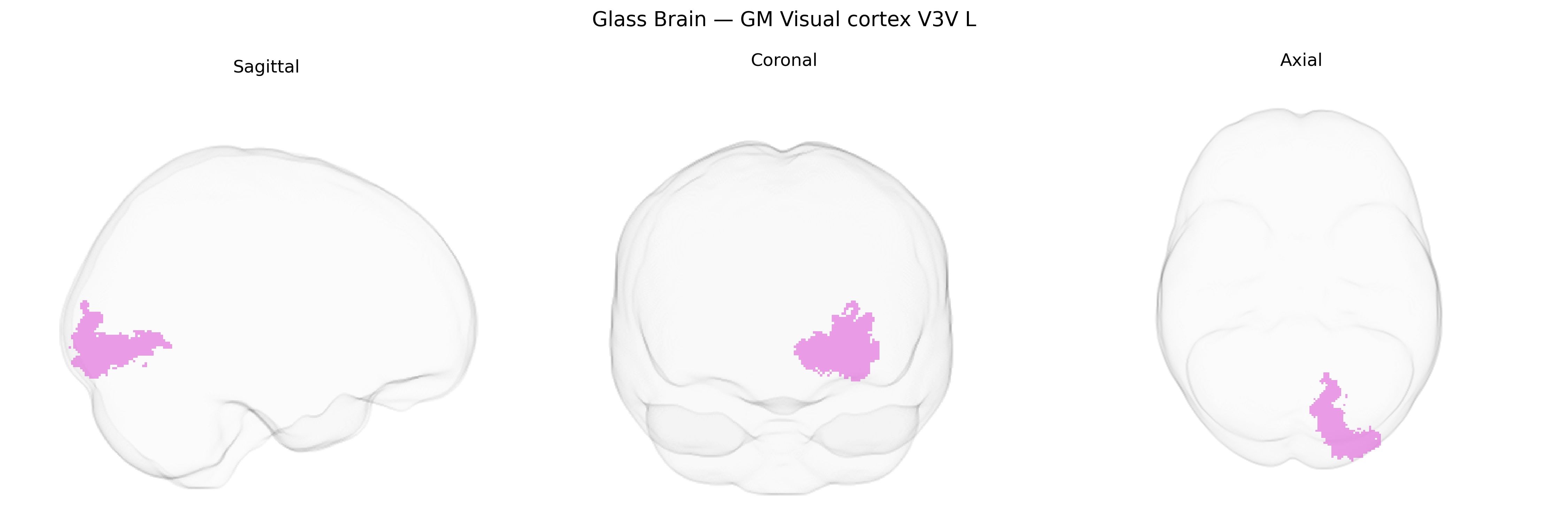
The bilateral GM Visual cortex V3V L region in the Juelich maxprob thr25 1mm atlas corresponds to the left ventral portion of visual area V3, situated in the occipital lobe along the ventral bank of the calcarine sulcus and adjacent to V2 and V4. This area is part of the early extrastriate visual cortex and contributes to the processing of form, orientation, and motion with a bias toward the lower visual field, integrating information within the ventral visual stream that supports object perception. Cytoarchitectonically, V3V shows distinct laminar organization relative to primary visual cortex (V1), with specialized neuronal populations and connectivity patterns that interface with both dorsal and ventral visual areas. There is no direct Wikipedia article specifically for “V3V”; a closely related and encompassing structure is Visual cortex.
The bilateral GM Visual cortex V3V L region from the Juelich maxprob thr25 1mm atlas, corresponding to ventral extrastriate visual area V3 (often overlapping with parts of the occipital and lateral occipital cortex), has been implicated in several genetic and GWAS-based findings primarily via large-scale imaging genetics studies rather than gene-to-region mappings specific to V3V. Twin and SNP-based heritability analyses show that gray-matter thickness, surface area, and functional activation in mid-level ventral visual areas, including V3/V3v, are substantially heritable, with polygenic influences distributed across many loci rather than a few region-specific genes. GWAS of occipital and visual-cortex cortical thickness and surface area (e.g., ENIGMA, UK Biobank) have reported associations with variants near genes involved in neurodevelopment, axon guidance, and synaptic function (such as variants in or near PAX6, FGFR3, TBR1/EMX2-related pathways, and multiple loci affecting global cortical morphology), though these findings generally map to broader occipital/visual regions rather than an anatomically isolated V3V L label. Genetic studies of visual disorders and traits that engage ventral visual cortex—including albinism, congenital prosopagnosia, and some forms of developmental dyslexia and autism—have identified risk genes influencing occipital and ventral stream structure and function, but these are typically described at the level of “occipital” or “ventral visual” cortex and not uniquely tied to V3V. Overall, current evidence supports robust but highly polygenic influences on the structure and function of this region, with genetic associations shared across neighboring visual areas rather than uniquely specific to the V3V L field as defined in the Juelich atlas.
Overview generated by GPT-4o (2026).
Region ID: 85
Hemisphere: bilateral
Atlas: Juelich maxprob thr25 1mm
Full Quality Version: Download MP4
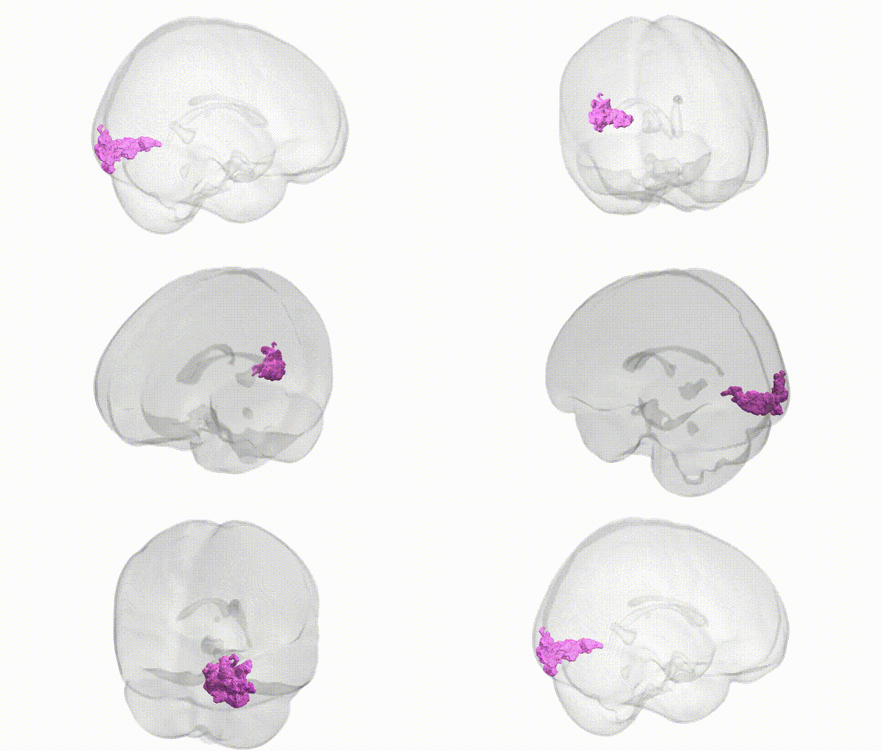
Full Quality Version: Download MP4
Wali Sidiqyar*, Gaurav Rudravaram*, Elyssa M. McMaster, Trent M. Schwartz, Adam M. Saunders, Kurt G. Schilling, Bennett A. Landman "Introducing SPINS: A Shared Public Visualization Library of Neuroanatomical Structures." Medical Imaging with Deep Learning- short paper
This resource is licensed under CC0 1.0 Universal (Public Domain).