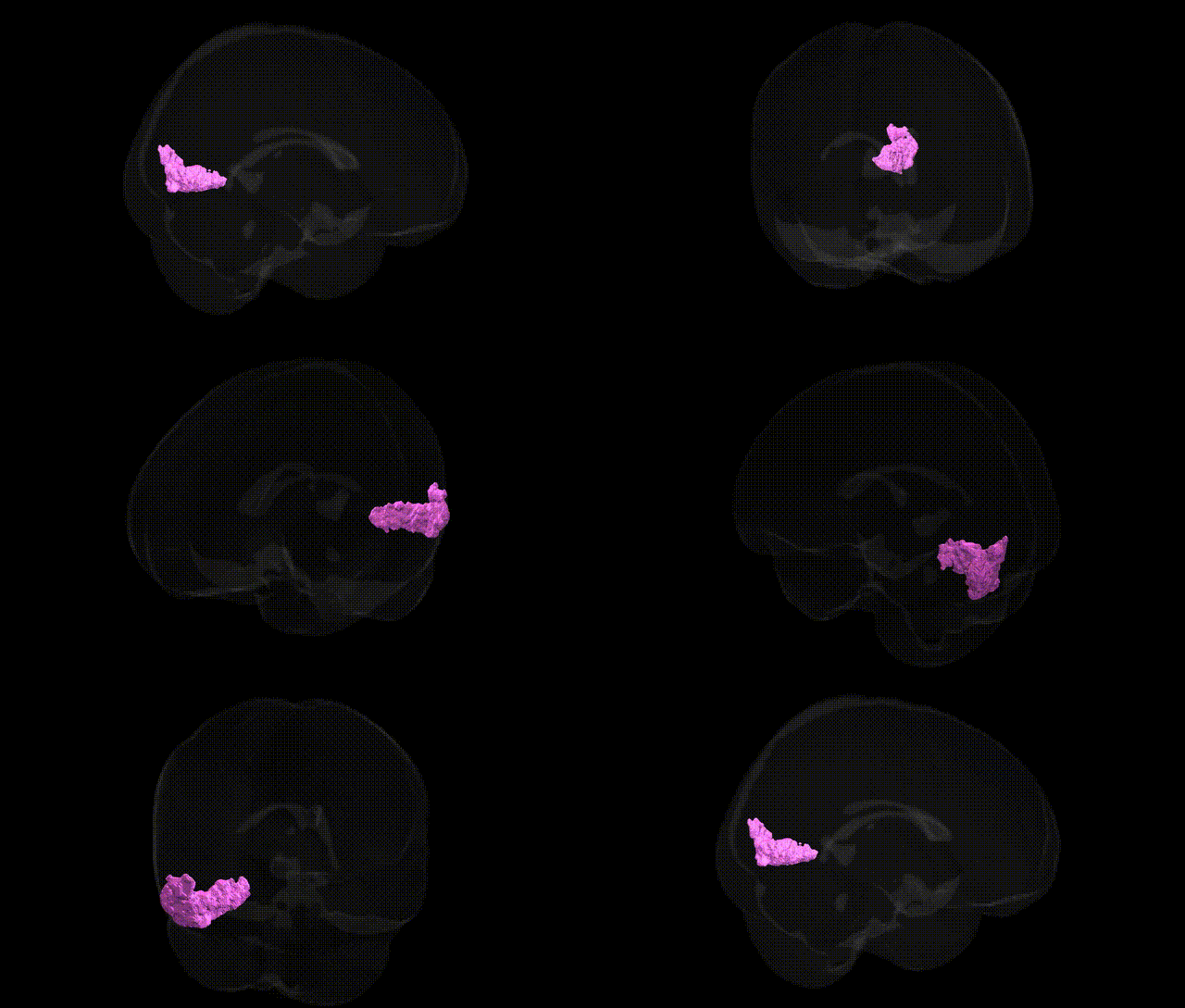
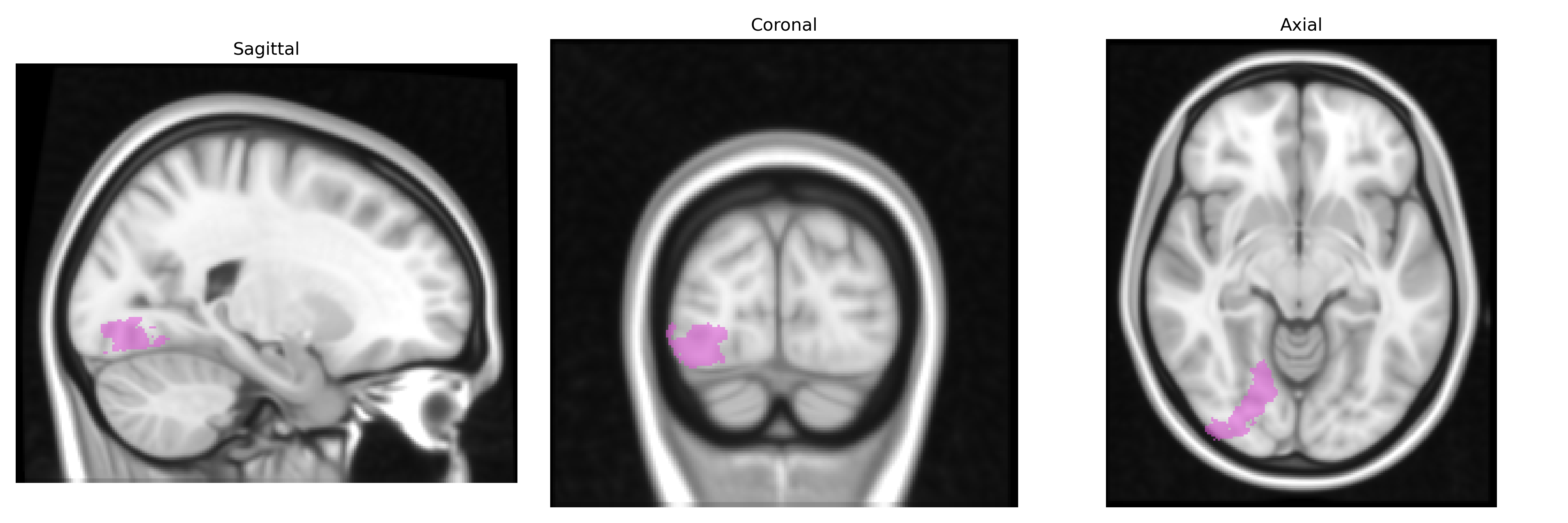
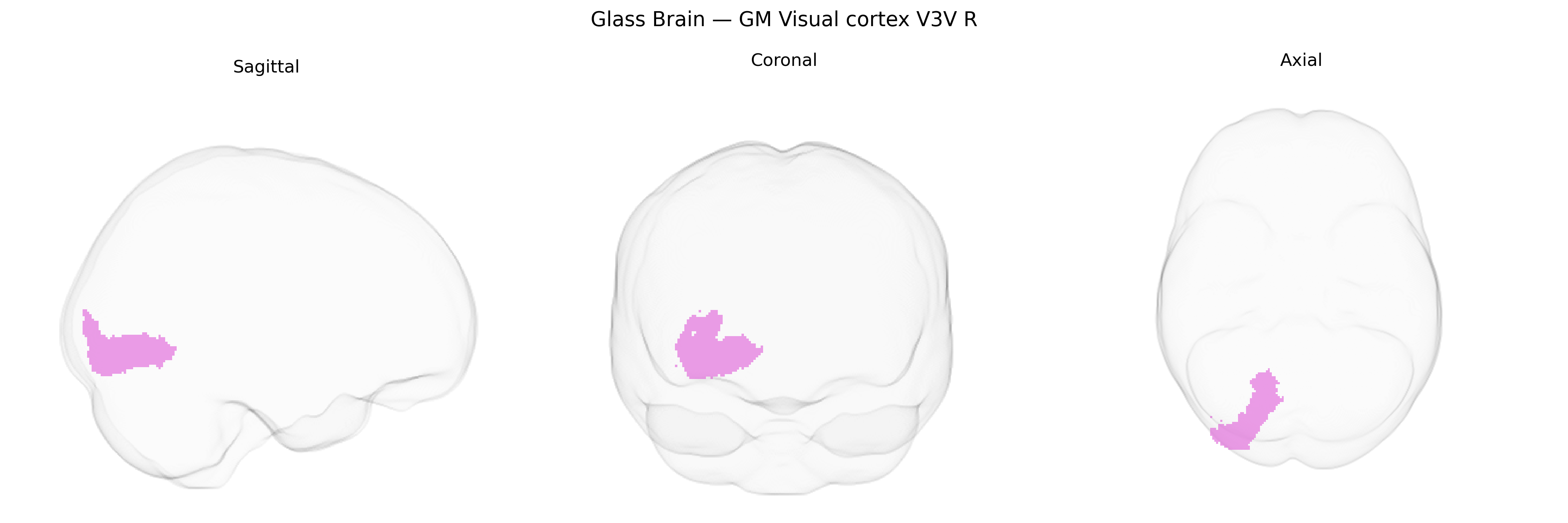
The bilateral GM Visual cortex V3V R region from the Juelich maxprob thr25 1mm Atlas corresponds to the ventral portion of visual area V3 in the right hemisphere, located in the occipital lobe along the ventral part of the dorsomedial visual cortex bordering areas V2 and V4. This region is part of the extrastriate visual cortex and contributes to intermediate-level visual processing, including analysis of form, orientation, and motion, with a stronger association to the ventral visual stream that supports object recognition. Cytoarchitectonically, V3V is characterized by distinct laminar organization typical of visual association areas, with prominent input from primary visual cortex (V1) and reciprocal connections to adjacent visual areas such as V2, V4, and higher-tier ventral stream regions. There is no direct Wikipedia article for V3V; a related and encompassing structure is the Visual cortex.
The bilateral GM Visual cortex V3V R region in the Juelich maxprob thr25 1mm Atlas corresponds to ventral extrastriate visual association cortex implicated in mid-level visual processing, and genetic associations identified for this territory come mainly from imaging–genetics and imaging–GWAS studies linking variants to occipital and ventral visual cortical thickness, surface area, and activation patterns. Large-scale GWAS of cortical structure (e.g., ENIGMA, UK Biobank) have reported that common variants in genes involved in neurodevelopment and synaptic function—such as variants near MEF2C, MAPT, and loci implicated in neuronal differentiation and axon guidance—are associated with structural metrics in occipital and ventral visual regions that include or overlap V3/V3v, although specific labeling as “V3V R” is rarely provided. Polygenic risk for schizophrenia, bipolar disorder, major depression, and autism spectrum disorder has been associated with altered occipital and ventral visual cortical morphology and functional responses, including regions encompassing V3, suggesting that shared genetic risk factors influencing synaptic plasticity, cortical layering, and visual network connectivity may contribute to vulnerability to these conditions. In addition, genetic variants linked to albinism, congenital visual pathway anomalies, and some forms of epilepsy and migraine show effects on occipital visual networks that likely involve V3V, but current GWAS and imaging–genetics literature does not yet define a distinct, region-specific genetic signature unique to the Juelich-defined bilateral V3V R area, with most findings reflecting broader visual cortical and occipito-temporal network associations.
Overview generated by GPT-4o (2026).
Region ID: 86
Hemisphere: bilateral
Atlas: Juelich maxprob thr25 1mm
Full Quality Version: Download MP4
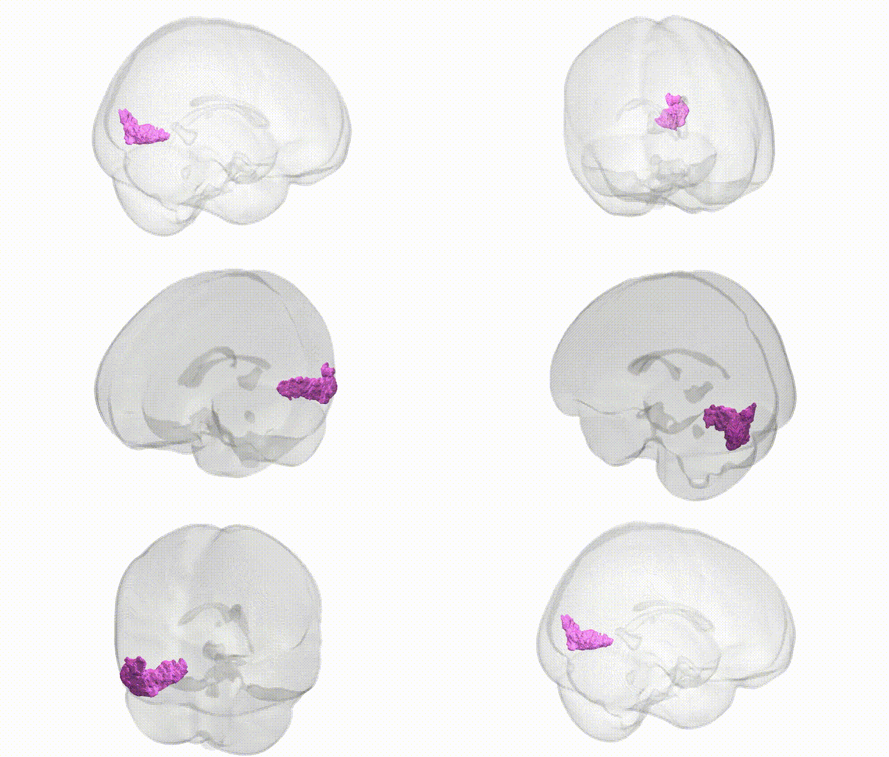
Full Quality Version: Download MP4
Wali Sidiqyar*, Gaurav Rudravaram*, Elyssa M. McMaster, Trent M. Schwartz, Adam M. Saunders, Kurt G. Schilling, Bennett A. Landman "Introducing SPINS: A Shared Public Visualization Library of Neuroanatomical Structures." Medical Imaging with Deep Learning- short paper
This resource is licensed under CC0 1.0 Universal (Public Domain).