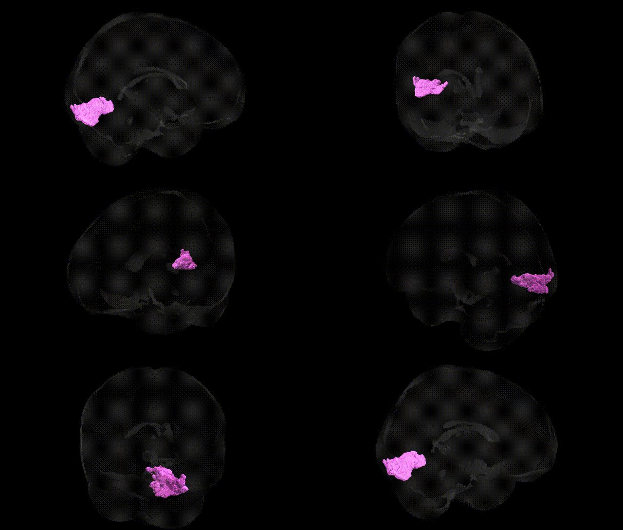
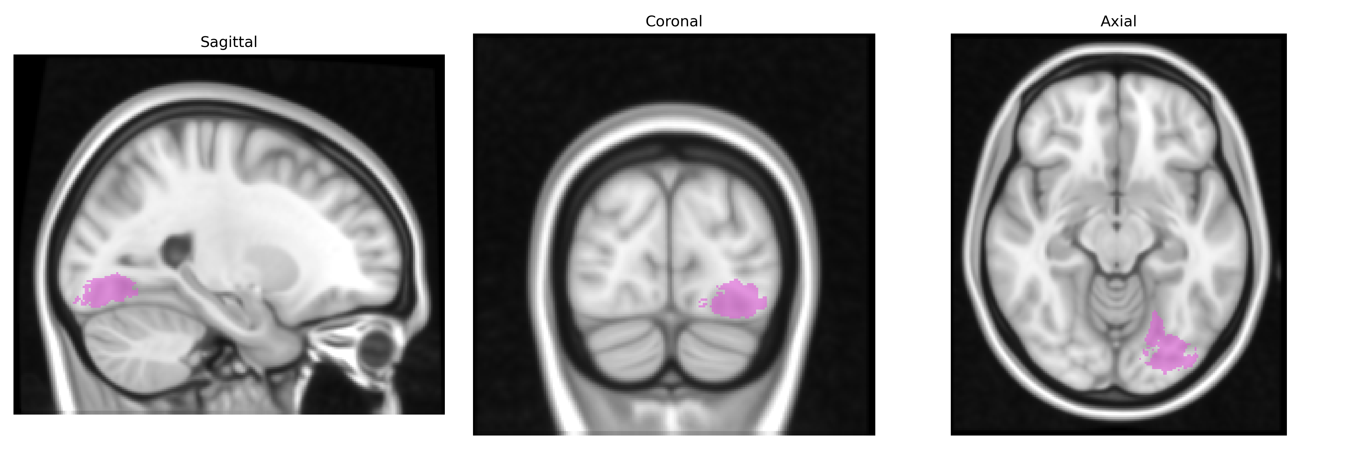
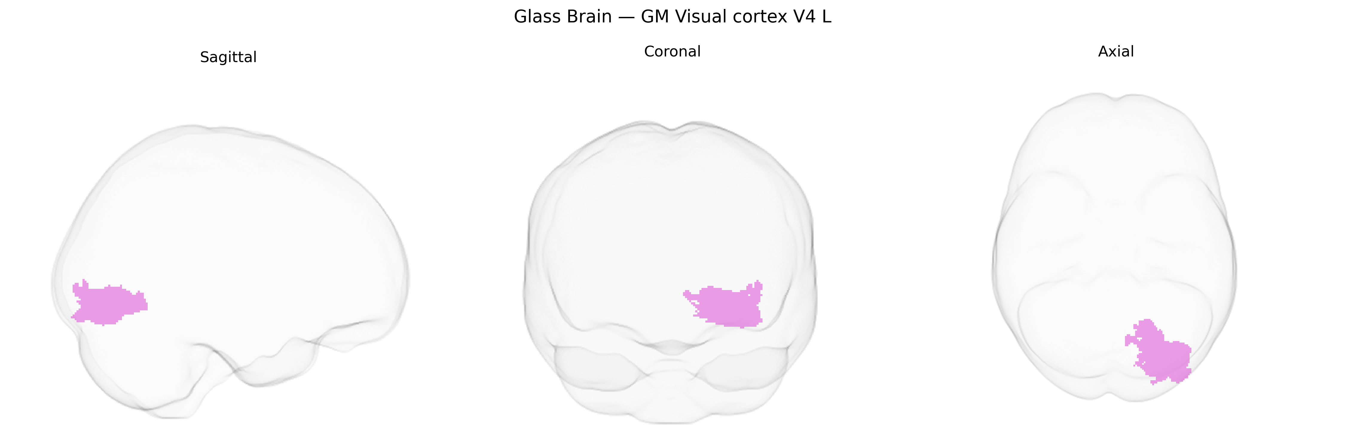
The bilateral GM Visual cortex V4 L region in the Juelich maxprob thr25 1mm Atlas corresponds to a left-hemispheric probabilistic map of human visual area V4, a mid-tier extrastriate visual area located on the ventral occipitotemporal cortex, typically along the fusiform and lingual gyri. This region is implicated in intermediate-level processing of visual information, including color perception, form and curvature processing, and contributes to the ventral “what” pathway for object recognition. Neurons in V4 exhibit selectivity for chromatic contrast, complex shapes, and contour features, and receive input from earlier visual areas such as V2 while projecting to higher-order ventral stream regions involved in object and category representations. As a defined human V4 entry is not available as a standalone article, a closely related structure is described under Visual cortex.
Bilateral GM visual cortex V4 (left hemisphere) from the Juelich maxprob thr25 1 mm atlas corresponds to a mid-level ventral visual area implicated in color processing, object recognition, and attention, but currently lacks robust, region-specific genetic associations from large-scale GWAS at the fine-grained atlas level. Most genetic findings involving this region are indirect, arising from imaging–genetics and imaging–GWAS studies that link common variants to occipital or ventral visual cortical thickness, surface area, or functional activation, rather than V4 specifically. Polygenic influences affecting general cortical development (e.g., variants near genes such as MAPT, TBR1, or those regulating synaptic plasticity and neurogenesis) have been associated with structural variation across occipital and temporal cortices, including regions encompassing V4, and visual association cortex measures appear in multivariate GWAS of cortical thickness and surface area. Disorders with strong genetic components that frequently show altered activation or structure in ventral visual areas—including autism spectrum disorder, schizophrenia, major depression, and certain forms of dyslexia—have implicated genes related to synaptic function (e.g., NRXN, NRGN, GRIN family), neurodevelopment, and neurotransmitter systems, but these associations are typically to broader networks (visual, attentional, or ventral stream circuits) rather than to V4 alone. Overall, current evidence indicates that genetic influences on V4L are part of distributed polygenic effects on visual association cortex, with no single variant or gene reproducibly and specifically tied to this Juelich-defined V4 region in large human GWAS to date.
Overview generated by GPT-4o (2026).
Region ID: 87
Hemisphere: bilateral
Atlas: Juelich maxprob thr25 1mm
Full Quality Version: Download MP4
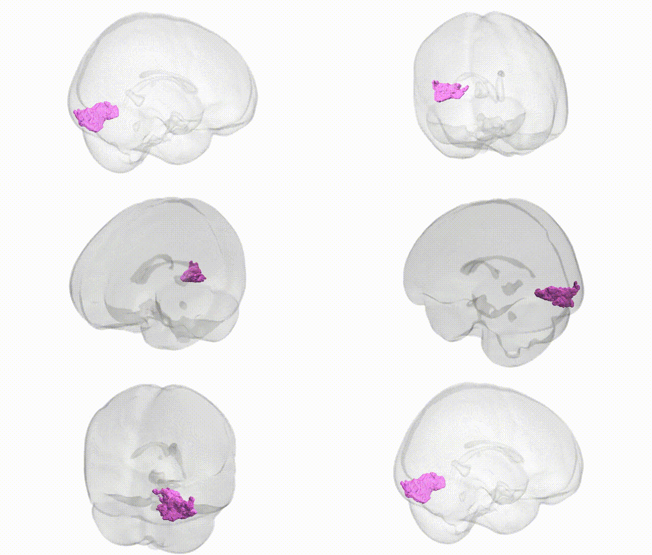
Full Quality Version: Download MP4
Wali Sidiqyar*, Gaurav Rudravaram*, Elyssa M. McMaster, Trent M. Schwartz, Adam M. Saunders, Kurt G. Schilling, Bennett A. Landman "Introducing SPINS: A Shared Public Visualization Library of Neuroanatomical Structures." Medical Imaging with Deep Learning- short paper
This resource is licensed under CC0 1.0 Universal (Public Domain).