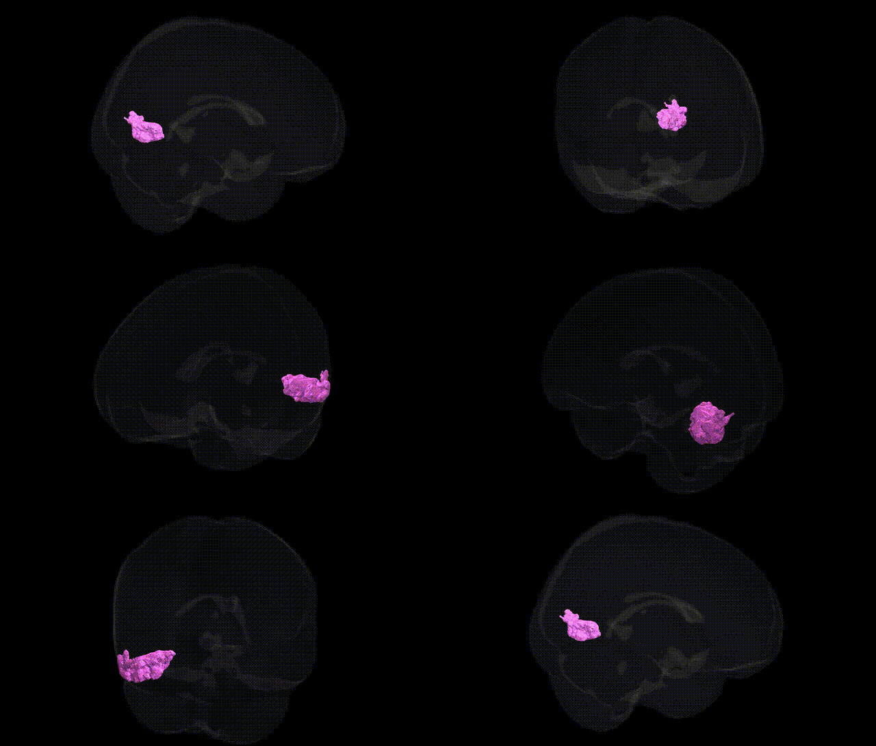
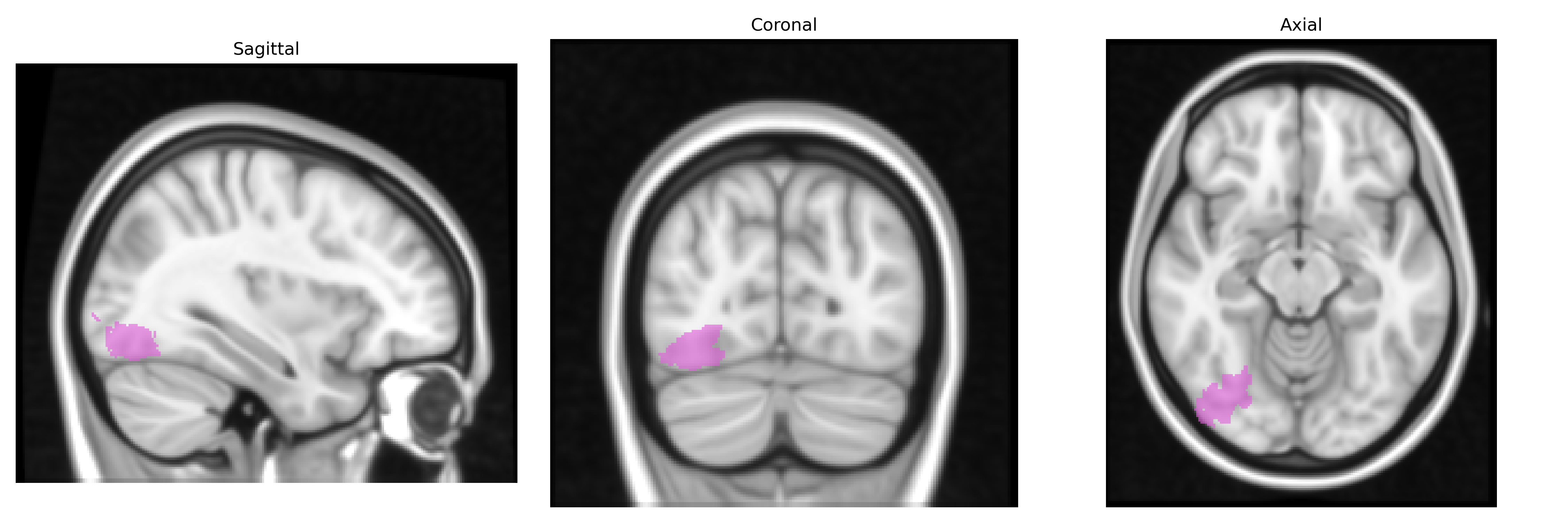
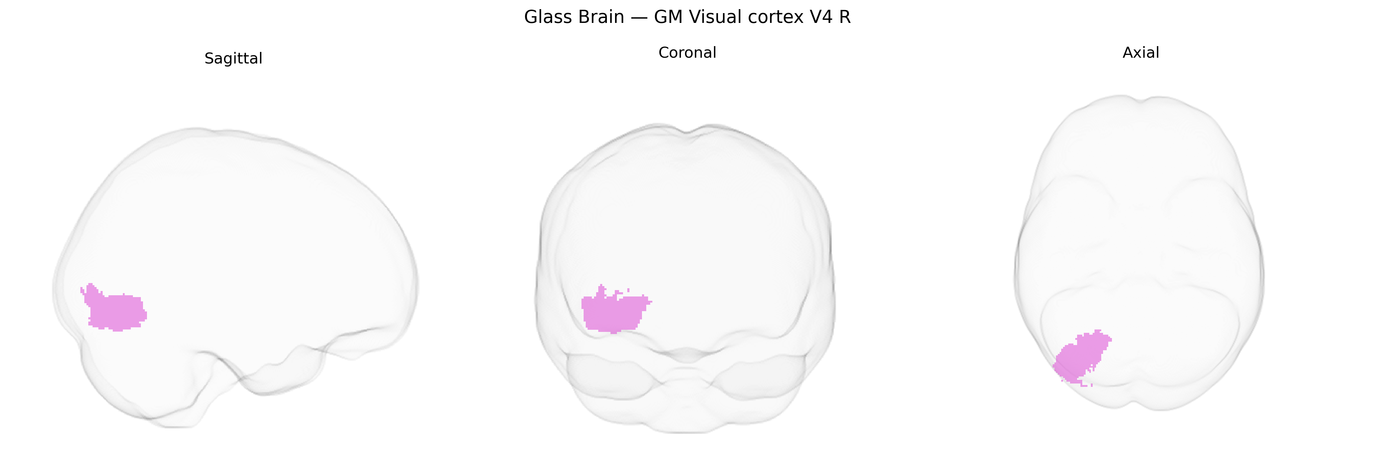
The bilateral GM Visual cortex V4 R region in the Juelich maxprob thr25 1mm Atlas corresponds to a right-hemispheric portion of area V4, a mid-tier extrastriate visual area located primarily on the ventral surface of the occipital lobe, extending into the occipito-temporal cortex. This region is implicated in intermediate-level visual processing, particularly in color perception, form and shape selectivity, and aspects of object recognition, and it receives substantial feedforward input from V2 and V3 while projecting to higher-order ventral visual stream regions such as inferotemporal cortex. Neurophysiological and neuroimaging studies indicate that V4 neurons exhibit sensitivity to chromatic contrast, curvature, and complex contour features, contributing critically to the integration of visual features into coherent object representations. There is no direct link for “V4 R,” but the closest related structure is Visual area V4.
The bilateral GM Visual cortex V4 R region in the Juelich maxprob thr25 1mm atlas corresponds to the mid-ventral occipitotemporal visual cortex involved in color processing, object recognition, and higher-order visual perception, and genetic associations are typically inferred from broader occipital/ventral visual regions rather than V4-specific GWAS signals. Large-scale imaging-genetics studies, such as those using UK Biobank data, have identified heritable variation in occipital and ventral visual cortical thickness and surface area linked to common variants near genes involved in neurodevelopment, axon guidance, and synaptic function (for example, loci near EOMES, TBR1, and other transcriptional regulators of corticogenesis), but these are usually reported at the level of occipital or visual association cortex rather than anatomically precise V4 fields. GWAS of visual cortical structure and function have associated variants in or near genes related to myelination and neuronal differentiation with differences in visual cortex volume and folding, and cross-trait analyses connect these loci to neurodevelopmental and psychiatric conditions (including autism spectrum disorder, schizophrenia, and major depression), suggesting that genetic factors influencing ventral visual cortex organization may partially overlap with those conferring risk for these disorders. In addition, candidate-gene and linkage studies in disorders with prominent visual-perceptual deficits—such as posterior cortical atrophy, certain familial forms of Alzheimer’s disease, and some monogenic visual processing disorders—implicate genes involved in amyloid processing, tau pathology, and synaptic stability, which can affect posterior cortical regions including V4, although these findings generally lack spatial specificity at the fine-grained cytoarchitectonic level. Overall, current evidence supports a polygenic architecture influencing the structure and function of ventral visual cortex encompassing V4, with genetic variants contributing to interindividual differences in visual perception and susceptibility to neurodevelopmental and neurodegenerative disorders, but no robust, V4-specific GWAS loci have yet been consistently isolated.
Overview generated by GPT-4o (2026).
Region ID: 88
Hemisphere: bilateral
Atlas: Juelich maxprob thr25 1mm
Full Quality Version: Download MP4
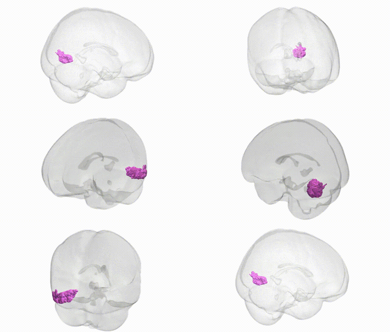
Full Quality Version: Download MP4
Wali Sidiqyar*, Gaurav Rudravaram*, Elyssa M. McMaster, Trent M. Schwartz, Adam M. Saunders, Kurt G. Schilling, Bennett A. Landman "Introducing SPINS: A Shared Public Visualization Library of Neuroanatomical Structures." Medical Imaging with Deep Learning- short paper
This resource is licensed under CC0 1.0 Universal (Public Domain).