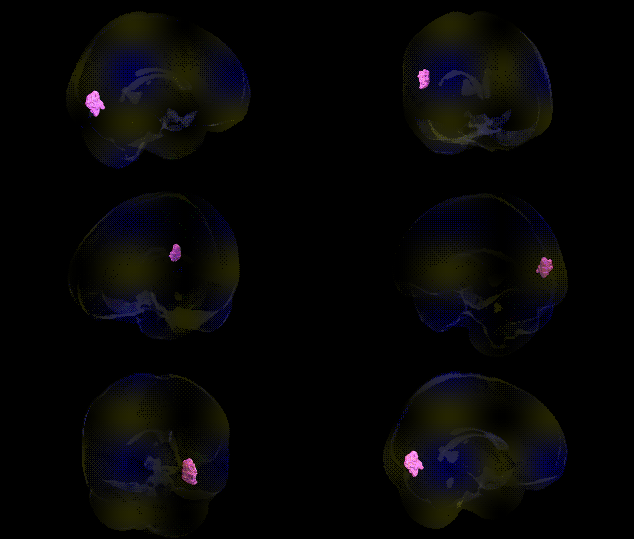
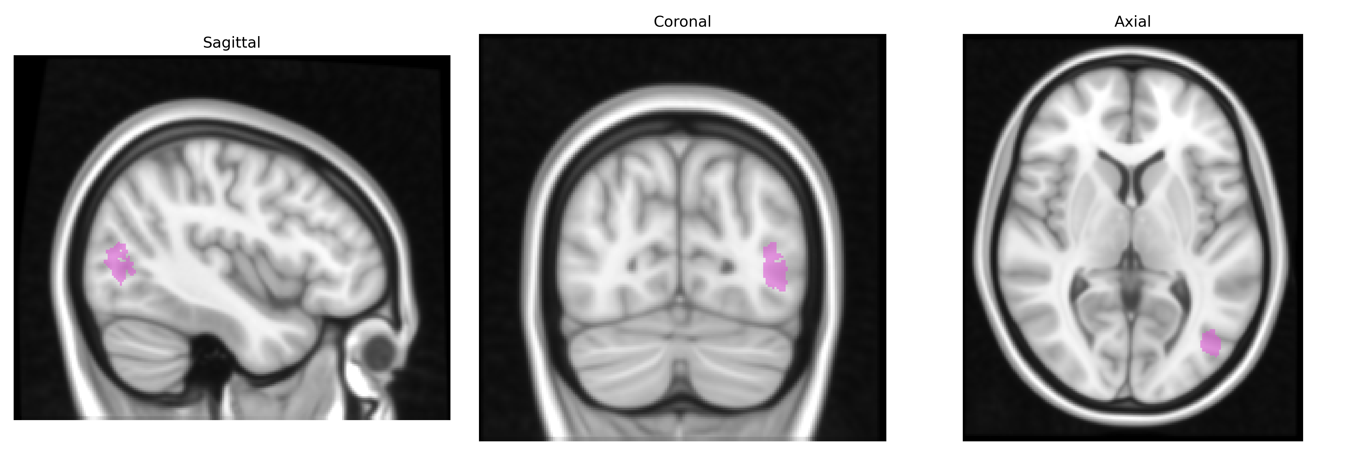
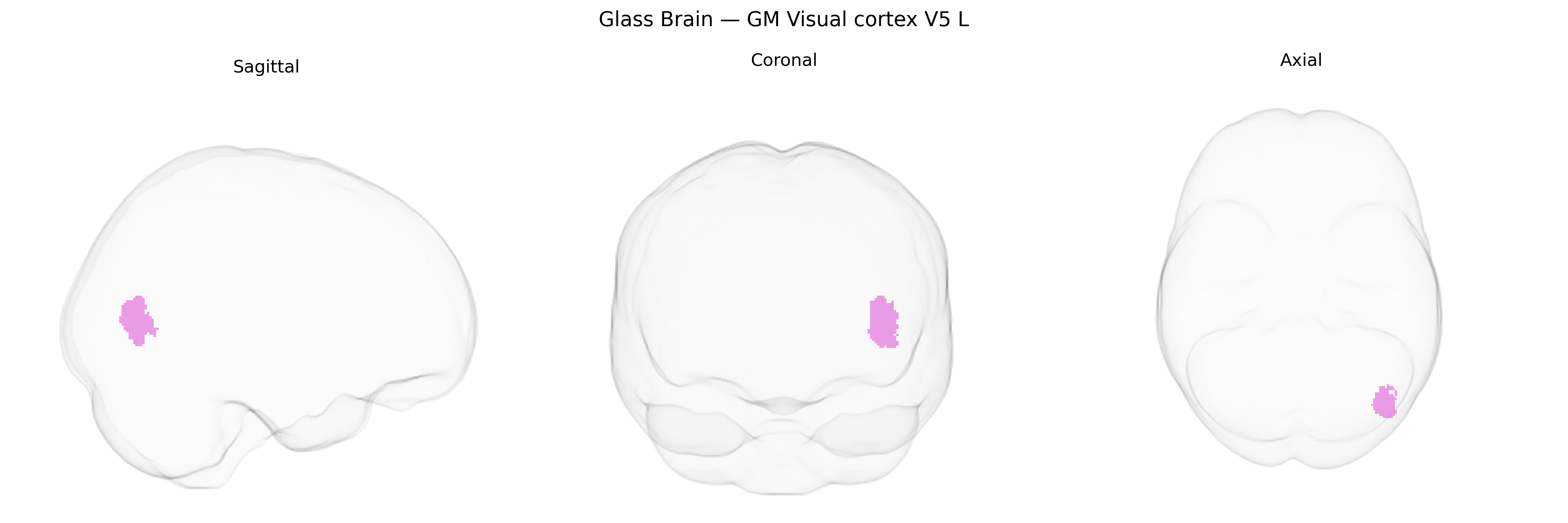
The bilateral GM Visual cortex V5 L region in the Juelich maxprob thr25 1mm atlas corresponds to the left-hemispheric portion of human area V5/MT, a motion-sensitive extrastriate visual area located in the lateral occipitotemporal cortex, typically along the ascending limb of the inferior temporal sulcus. This region is characterized by strong direction-selective responses to moving stimuli, sensitivity to motion coherence, and involvement in the perception of global motion, optic flow, and motion-defined form. Cytoarchitectonically, it is distinct from neighboring visual areas such as V3 and V4, and it forms part of the dorsal visual processing stream, projecting to parietal regions involved in visuomotor integration and spatial attention. Lesions in this area can give rise to motion perception deficits such as akinetopsia. No direct Wikipedia article exists for this exact parcellation; a closely related structure is described at V5.
The bilateral GM Visual cortex V5 (MT) L region in the Juelich maxprob thr25 1mm atlas corresponds to motion-sensitive extrastriate cortex, which has been implicated in genetic studies of visual processing, cortical structure, and neuropsychiatric risk. Large-scale imaging–genetics GWAS (e.g., ENIGMA, UK Biobank) have identified common variants associated with occipital and temporo-occipital cortical thickness and surface area, frequently involving loci near genes such as PAX6, TCF4, MEF2C, and other regulators of neurodevelopment and synaptic function, though these associations are typically reported at lobar or broader regional levels rather than specifically labeled V5. Polygenic risk scores for schizophrenia, bipolar disorder, and major depression have been linked to structural and functional alterations in extrastriate visual regions, including MT/V5, with some studies indicating that genetic liability to psychosis correlates with reduced gray matter or altered activation in motion-processing cortex. Variants in genes related to myelination and visual pathway development (e.g., those near CNTNAP2, NRXN1) have been associated with white matter integrity in optic radiations and dorsal visual stream tracts that project to MT/V5, which may indirectly influence this region. Additionally, GWAS of brain functional networks have implicated genetic effects on dorsal visual and attention networks that encompass V5/MT, connecting this region to heritable differences in visuospatial attention, motion perception, and reading-related traits; however, no single gene or variant is currently established as a specific, robust determinant of V5 structure or function, and most evidence comes from broader occipital or network-level imaging–genetics analyses rather than region-specific GWAS targeting Juelich-defined V5.
Overview generated by GPT-4o (2026).
Region ID: 89
Hemisphere: bilateral
Atlas: Juelich maxprob thr25 1mm
Full Quality Version: Download MP4
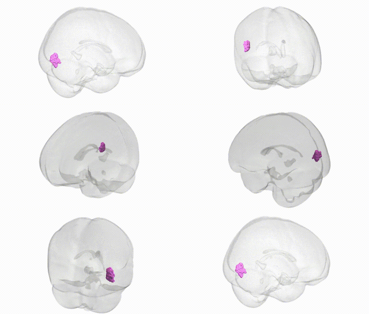
Full Quality Version: Download MP4
Wali Sidiqyar*, Gaurav Rudravaram*, Elyssa M. McMaster, Trent M. Schwartz, Adam M. Saunders, Kurt G. Schilling, Bennett A. Landman "Introducing SPINS: A Shared Public Visualization Library of Neuroanatomical Structures." Medical Imaging with Deep Learning- short paper
This resource is licensed under CC0 1.0 Universal (Public Domain).