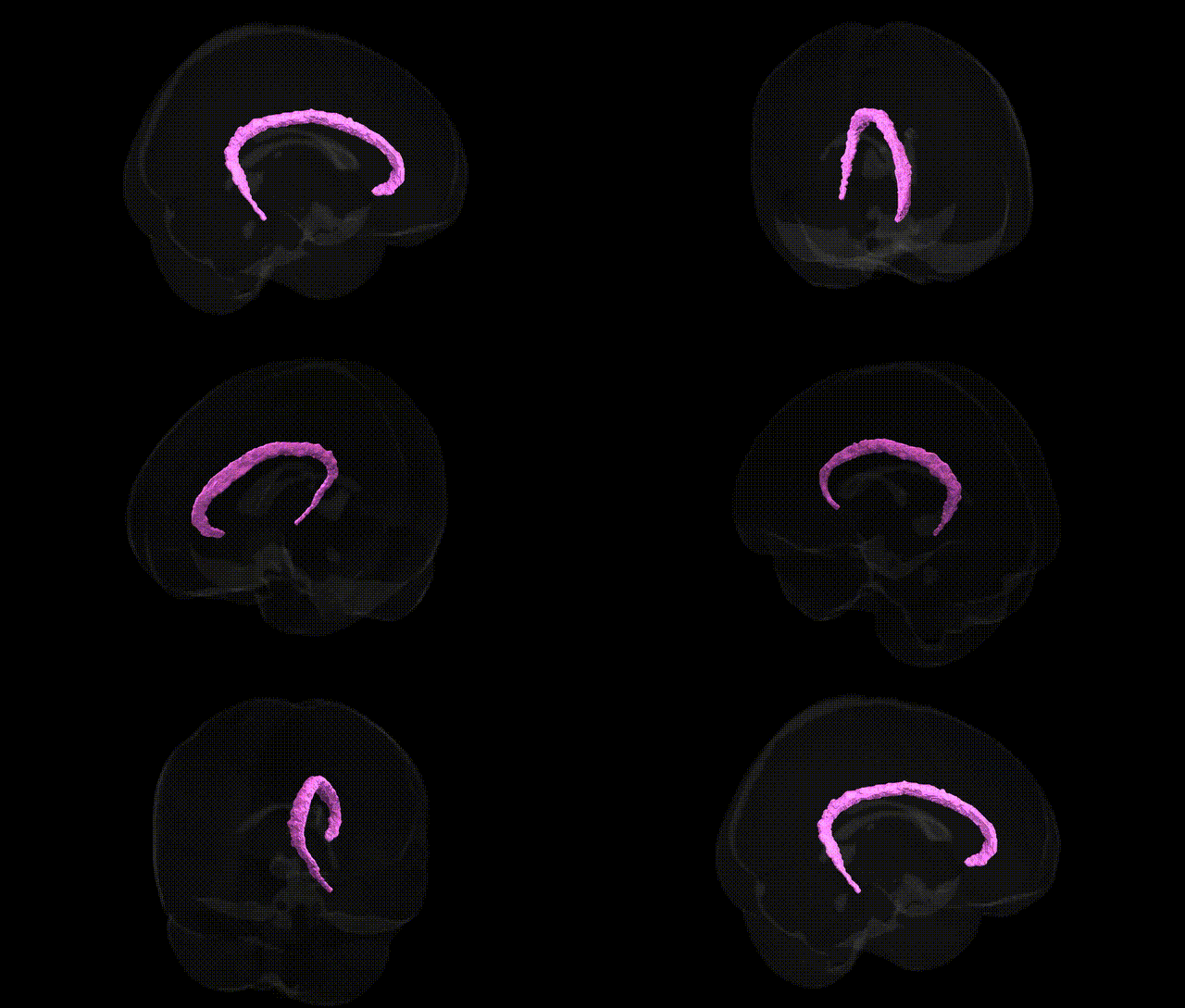
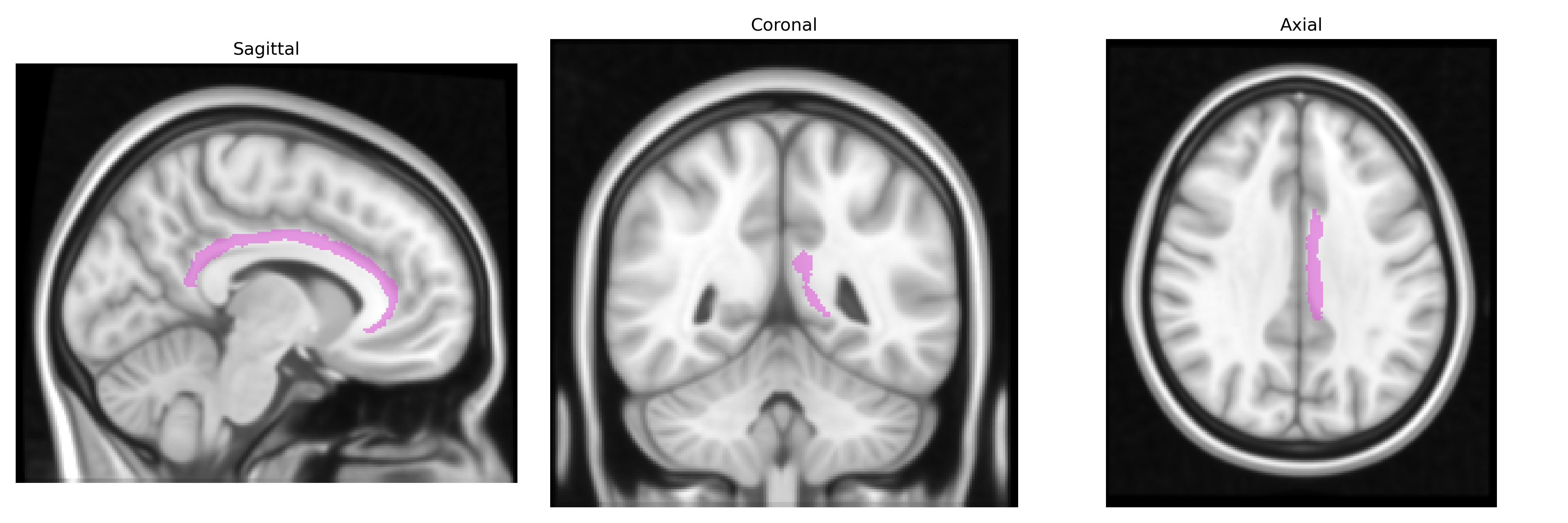
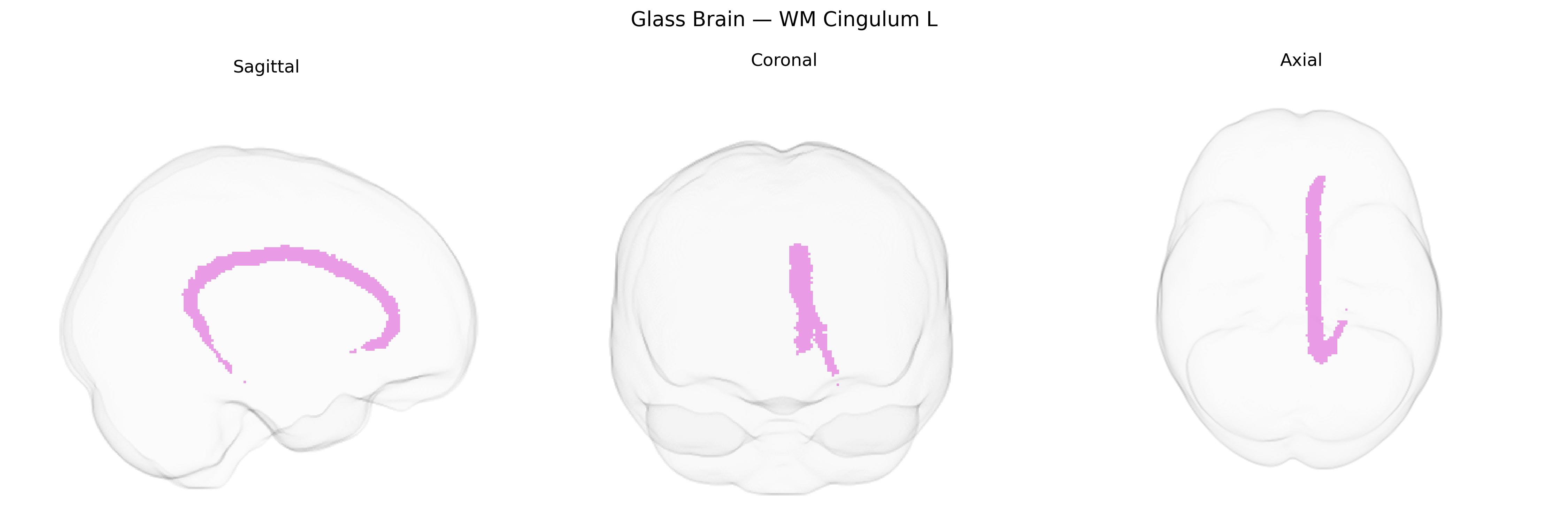
The bilateral WM Cingulum L in the Juelich maxprob thr25 1mm atlas refers to the left cingulum white matter bundle, a major association tract coursing within the cingulate gyrus and extending through the cingulate and parahippocampal regions. This tract interconnects medial frontal, cingulate, and medial temporal structures, including components of the limbic system, and plays a central role in integrating cognitive, emotional, and mnemonic processes. Anatomically, it runs parallel to the corpus callosum, arching around it and linking areas such as the anterior cingulate cortex, posterior cingulate cortex, and parahippocampal gyrus, thereby supporting functions related to attention, executive control, and episodic memory. There is no direct Wikipedia article for “bilateral WM Cingulum L”; a related structure is the Cingulum (brain).
The bilateral white matter cingulum (left) from the Juelich maxprob thr25 1 mm atlas lies within the core structural pathway of the cingulate/limbic system, and genetic associations typically emerge from studies of cingulum microstructure (e.g., fractional anisotropy) rather than regional labels per se. Twin and imaging-genetics studies indicate high heritability of cingulum integrity, with common variants in genes related to myelination, axon guidance, and synaptic plasticity (such as NTRK1/2, BDNF, and oligodendrocyte-related genes) repeatedly implicated, though often not at genome‑wide significance in region‑specific analyses. Large GWAS of white matter diffusion metrics (e.g., ENIGMA, UK Biobank) have reported associations between cingulum tract measures and loci in neurodevelopmental and cell-adhesion genes (including regions near CNTN4/CNTN6, LINGO1, and MAG), suggesting that genetic variation affecting axonal growth and myelin maintenance influences this tract. Disorder‑related imaging-genetics work has linked cingulum microstructural alterations to schizophrenia, major depressive disorder, bipolar disorder, ADHD, and PTSD, with risk alleles in genes such as CACNA1C, ZNF804A, and serotonin-related loci showing secondary associations with cingulum diffusion metrics or cingulate connectivity, although these findings are often indirect, trait‑mediated, and not specific to the left cingulum segment in the Juelich atlas. Overall, the genetic signal for this region reflects broad neurodevelopmental and myelination pathways rather than discrete, well‑replicated, region‑specific GWAS hits.
Overview generated by GPT-4o (2026).
Region ID: 97
Hemisphere: bilateral
Atlas: Juelich maxprob thr25 1mm
Full Quality Version: Download MP4
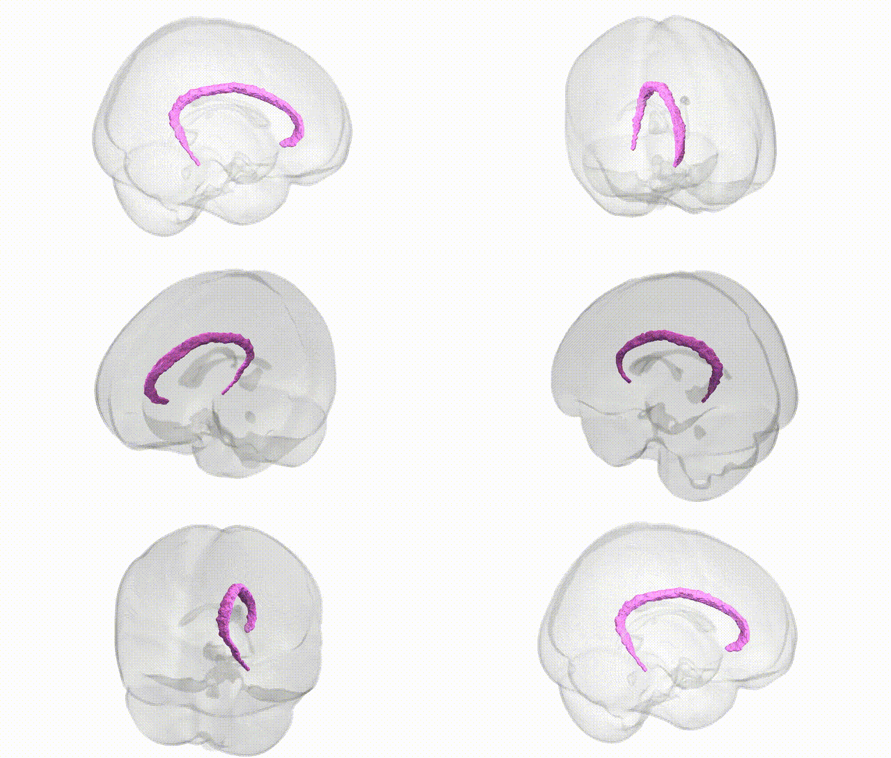
Full Quality Version: Download MP4
Wali Sidiqyar*, Gaurav Rudravaram*, Elyssa M. McMaster, Trent M. Schwartz, Adam M. Saunders, Kurt G. Schilling, Bennett A. Landman "Introducing SPINS: A Shared Public Visualization Library of Neuroanatomical Structures." Medical Imaging with Deep Learning- short paper
This resource is licensed under CC0 1.0 Universal (Public Domain).