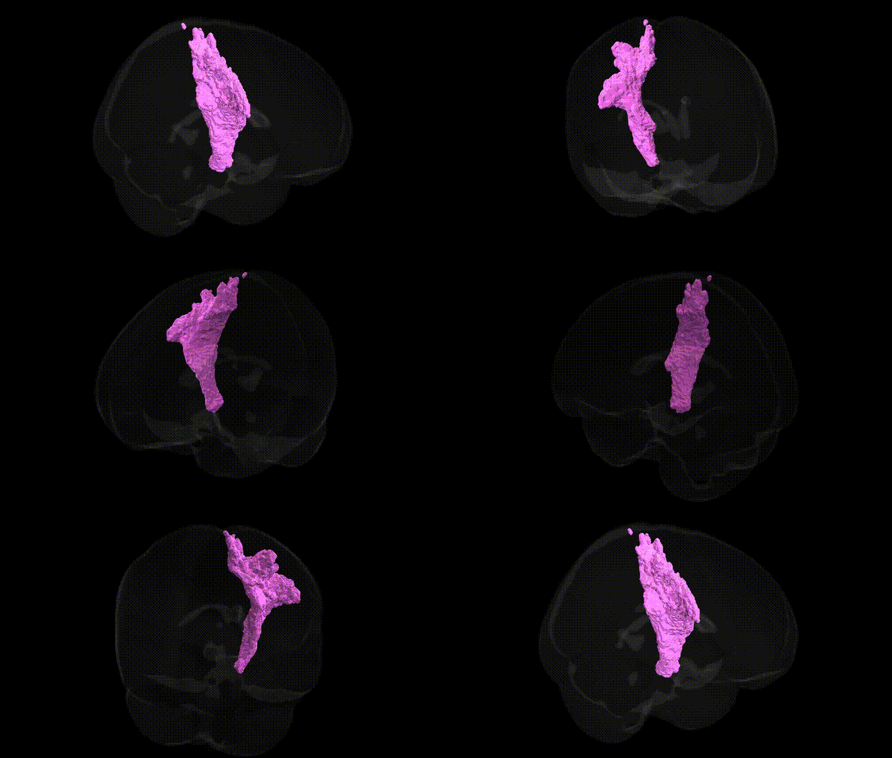
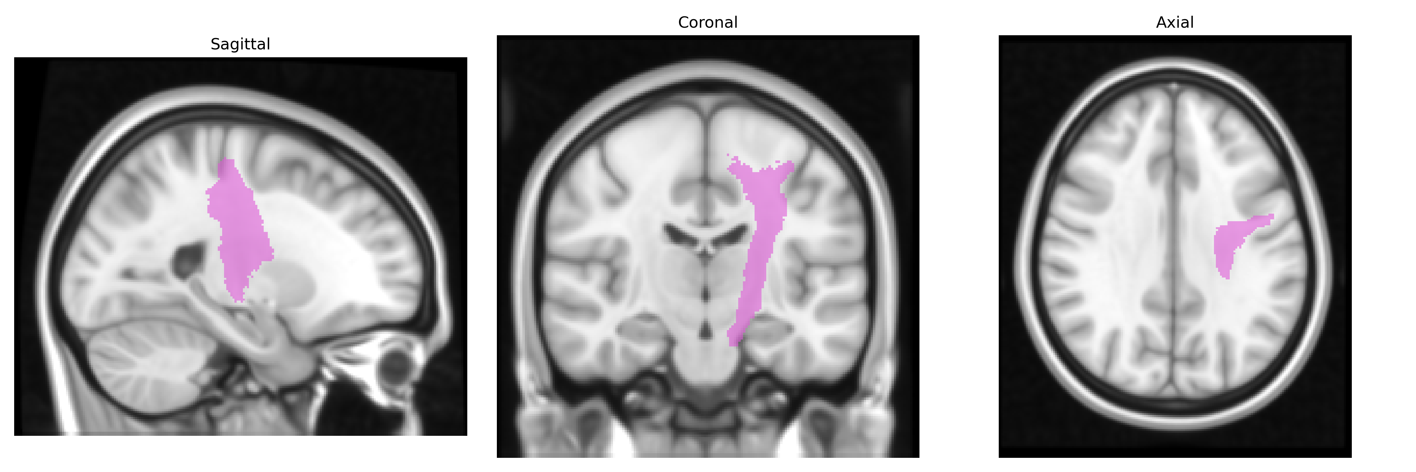
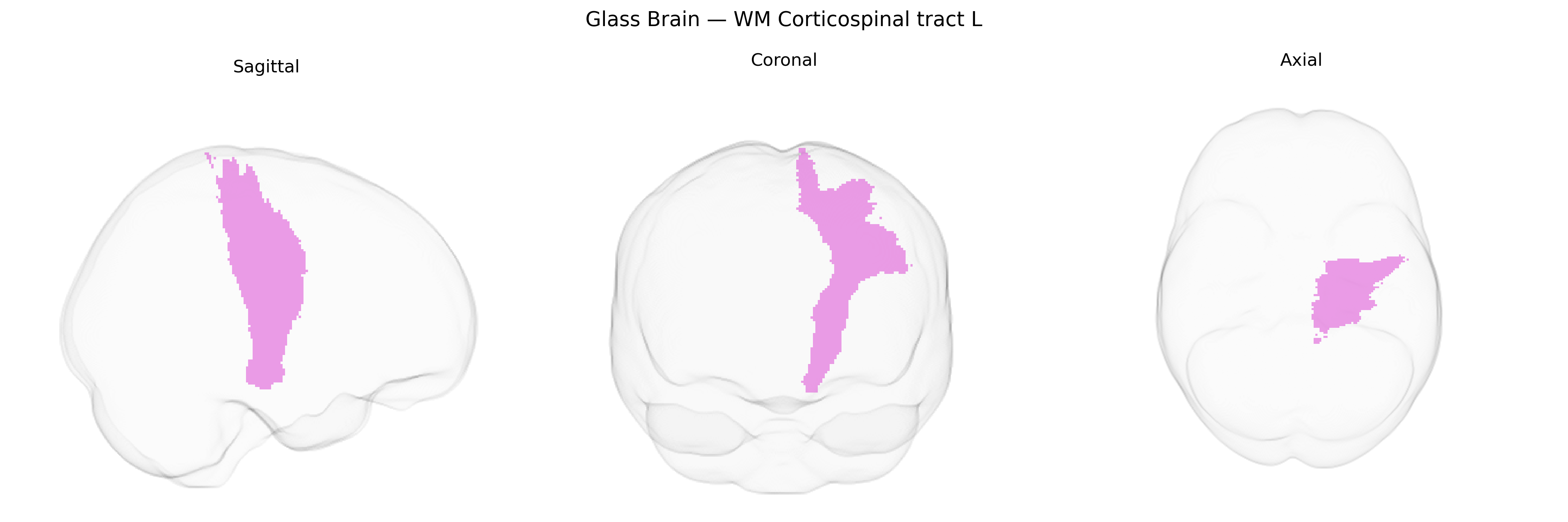
The bilateral WM Corticospinal tract L, as defined in the Juelich maxprob thr25 1 mm atlas, corresponds to left-hemispheric white matter portions of the corticospinal pathway, a major descending motor tract that originates primarily from pyramidal neurons in the primary motor cortex, premotor areas, and supplementary motor areas. These fibers descend through the corona radiata and posterior limb of the internal capsule, traverse the cerebral peduncles in the midbrain, continue through the ventral pons, and form the medullary pyramids before most fibers decussate at the pyramidal decussation in the caudal medulla to innervate contralateral spinal motor circuits. Functionally, this tract is critical for the execution and fine control of voluntary, especially distal, movements of the limbs and digits, mediating fast, precise activation of alpha motor neurons and interneurons in the spinal cord. While there is no dedicated Wikipedia page for the specific atlas-defined “bilateral WM Corticospinal tract L” region, it is a component of the broader Corticospinal tract.
The bilateral white-matter corticospinal tract in the left hemisphere, as defined in the Juelich maxprob thr25 1 mm atlas, has not been the primary focus of region-specific GWAS, but genetic studies of corticospinal integrity and motor-system white matter implicate polygenic influences related to axon guidance, myelination, and neuroinflammation. Variants in genes such as CNTNAP2, NRG1, ERBB4, and those within major histocompatibility complex and immune-related pathways have been associated with white-matter microstructure metrics (e.g., fractional anisotropy) in large diffusion MRI GWAS, which include the corticospinal tract. Disorders strongly involving corticospinal pathways—amyotrophic lateral sclerosis, primary lateral sclerosis, hereditary spastic paraplegias, and certain forms of cerebral palsy—show convergent genetic associations in genes related to motor neuron survival (e.g., SOD1, C9orf72), axonal transport (e.g., KIF5A, SPAST), mitochondrial function, and synaptic regulation, with imaging-genetic studies linking these risk variants to corticospinal tract degeneration or altered microstructure. Additional large-scale imaging-genetics consortia (e.g., ENIGMA, UK Biobank analyses) have reported that polygenic risk scores for neurodevelopmental and psychiatric disorders (including schizophrenia, ADHD, and autism spectrum disorder) correlate with widespread white-matter changes that encompass the corticospinal tract, although no single variant or locus is uniquely specific to the left corticospinal tract in current literature.
Overview generated by GPT-4o (2026).
Region ID: 99
Hemisphere: bilateral
Atlas: Juelich maxprob thr25 1mm
Full Quality Version: Download MP4
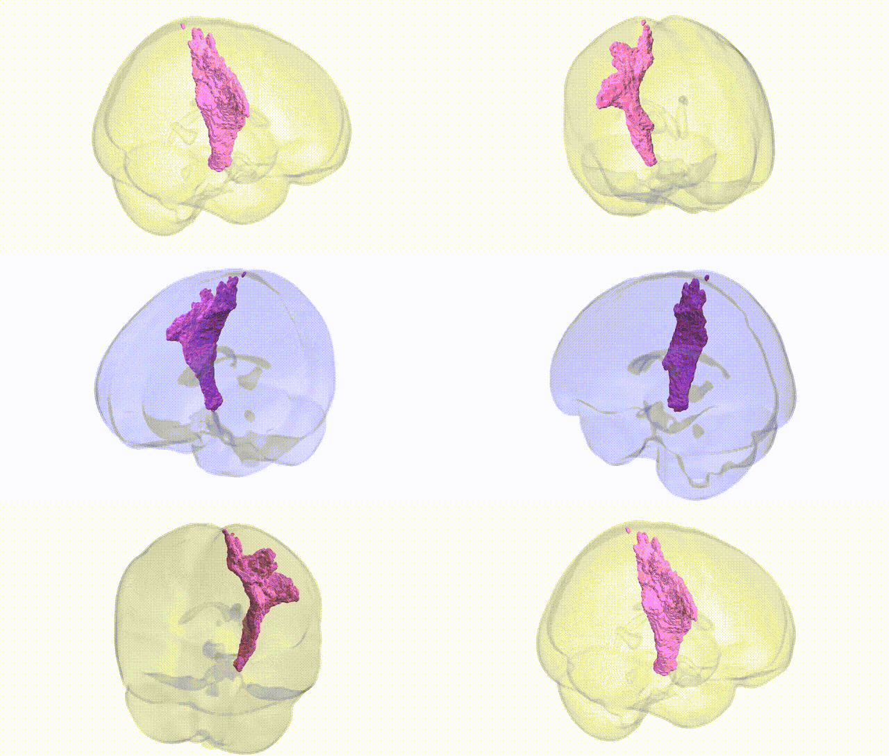
Full Quality Version: Download MP4
Wali Sidiqyar*, Gaurav Rudravaram*, Elyssa M. McMaster, Trent M. Schwartz, Adam M. Saunders, Kurt G. Schilling, Bennett A. Landman "Introducing SPINS: A Shared Public Visualization Library of Neuroanatomical Structures." Medical Imaging with Deep Learning- short paper
This resource is licensed under CC0 1.0 Universal (Public Domain).