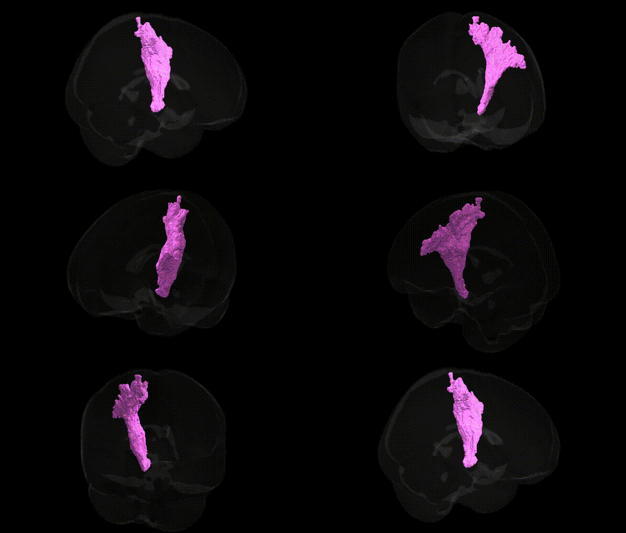
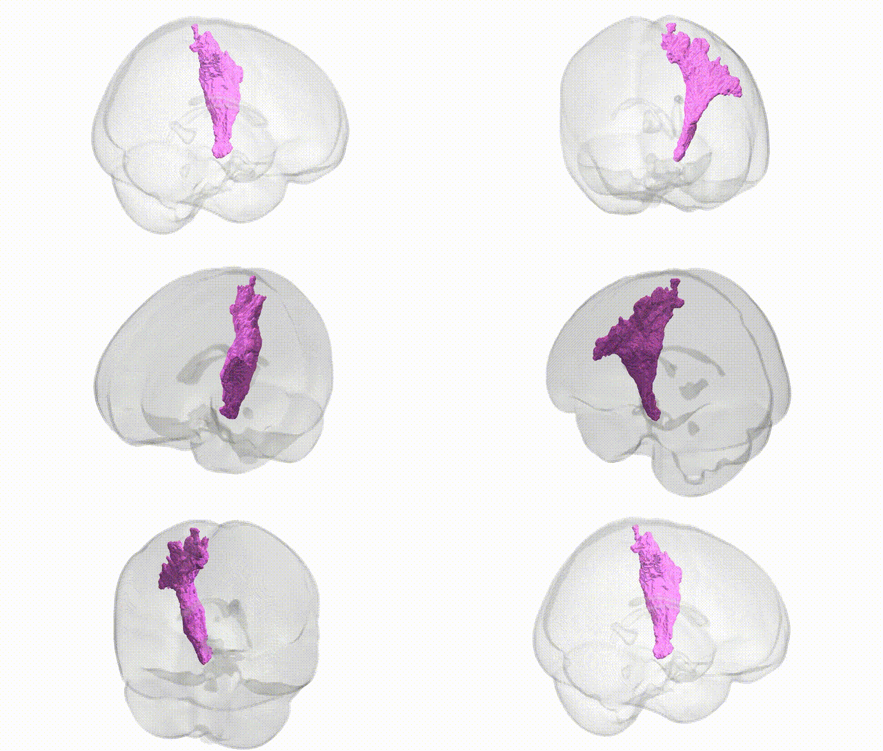
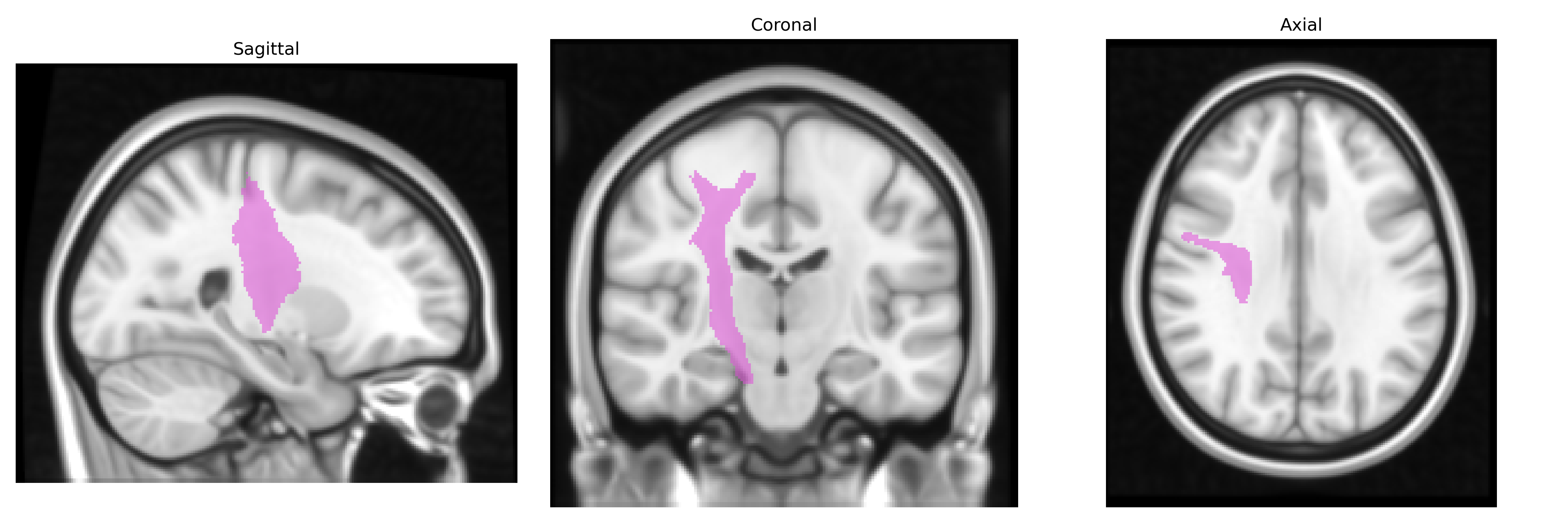
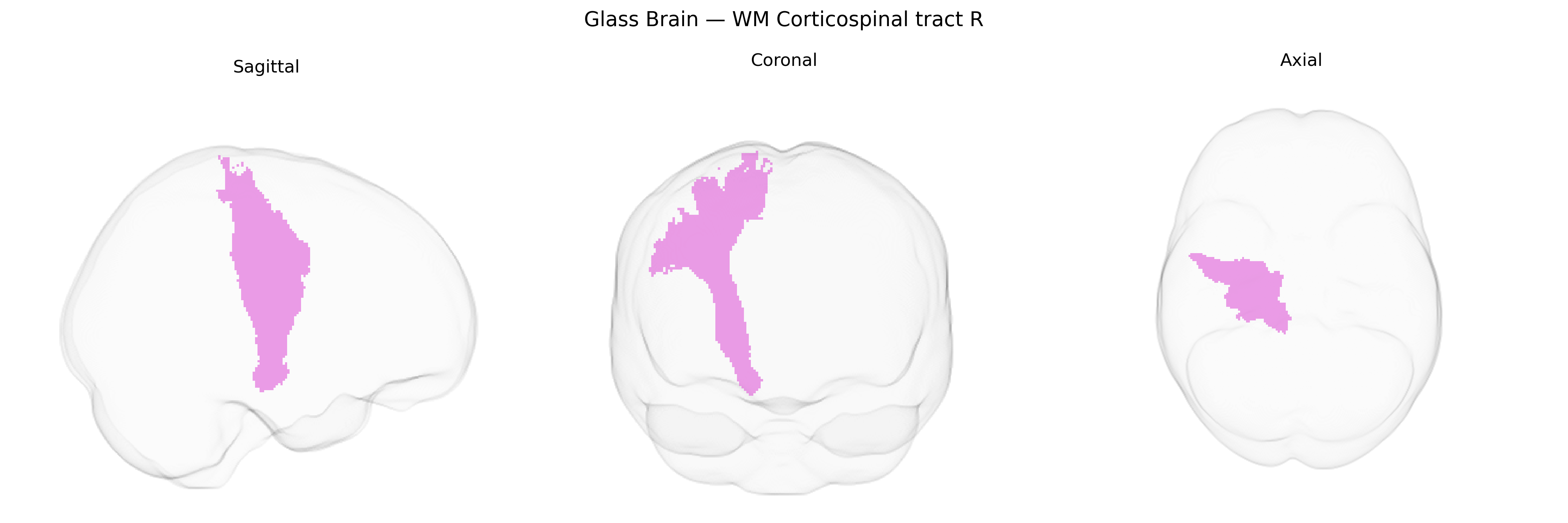
The bilateral WM Corticospinal tract R, as defined in the Juelich maxprob thr25 1mm atlas, corresponds to the right-lateralized portion of the corticospinal white matter pathway that conveys fast, primarily excitatory motor commands from the cerebral cortex to the spinal cord. Originating mainly from pyramidal neurons in primary motor, premotor, and supplementary motor cortices, its fibers descend through the corona radiata, internal capsule, cerebral peduncle, pons, and medullary pyramids before projecting to spinal motor circuits that control voluntary movement, particularly of distal limb musculature. This tract is essential for fine motor control, shows somatotopic organization, and is a major substrate for motor deficits following focal lesions such as stroke or traumatic injury. There is no direct Wikipedia article for this specific atlas label; a closely related structure with detailed information is the Corticospinal tract.
The right corticospinal tract white matter, as defined in the Juelich maxprob thr25 1 mm atlas, has been implicated indirectly in genetic studies through its microstructural properties and involvement in motor and neuropsychiatric phenotypes rather than via region-specific single-gene associations. Large-scale GWAS of diffusion MRI measures (e.g., fractional anisotropy and mean diffusivity) have identified multiple common variants—often in genes involved in axon guidance, myelination, and neuronal development (such as ROBO1/2, NRG1/ERBB signaling components, and oligodendrocyte-related genes)—that influence white matter integrity in major motor pathways including the corticospinal tract. Polygenic architectures associated with neurodevelopmental and neuropsychiatric disorders (e.g., schizophrenia, ADHD, autism) and neurodegenerative conditions (e.g., amyotrophic lateral sclerosis, multiple sclerosis) show convergent effects on corticospinal tract structure or connectivity in imaging–genetics and imaging–GWAS studies, although the associations are generally nonspecific to the right tract and often bilateral or global. In ALS, for example, risk variants in genes such as C9orf72, SOD1, and TARDBP are linked, via imaging, to degeneration and reduced integrity of corticospinal pathways, while MS risk loci in immune-related genes (e.g., HLA-DRB1*15:01) are associated with lesion burden and demyelination that frequently involve corticospinal fibers. Overall, genetic influences on the bilateral corticospinal tract R region are best characterized at the level of polygenic effects on white matter microstructure and motor-system vulnerability, rather than single, tract-specific genetic variants.
Overview generated by GPT-4o (2026).
Region ID: 98
Hemisphere: bilateral
Atlas: Juelich maxprob thr25 1mm
Full Quality Version: Download MP4
Full Quality Version: Download MP4
Wali Sidiqyar*, Gaurav Rudravaram*, Elyssa M. McMaster, Trent M. Schwartz, Adam M. Saunders, Kurt G. Schilling, Bennett A. Landman "Introducing SPINS: A Shared Public Visualization Library of Neuroanatomical Structures." Medical Imaging with Deep Learning- short paper
This resource is licensed under CC0 1.0 Universal (Public Domain).