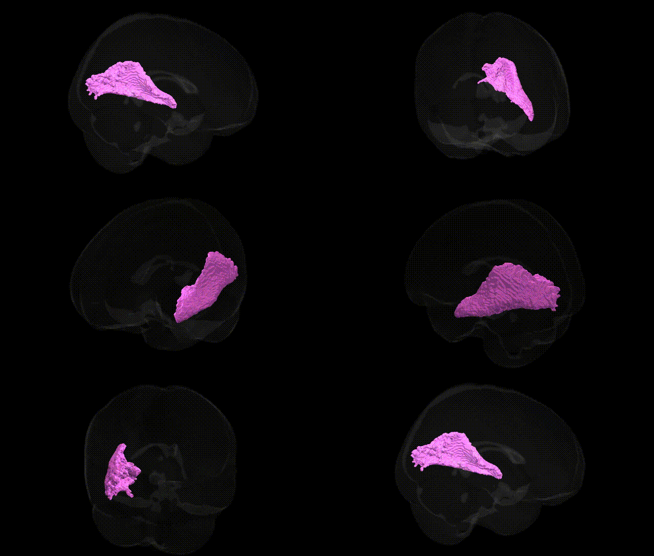
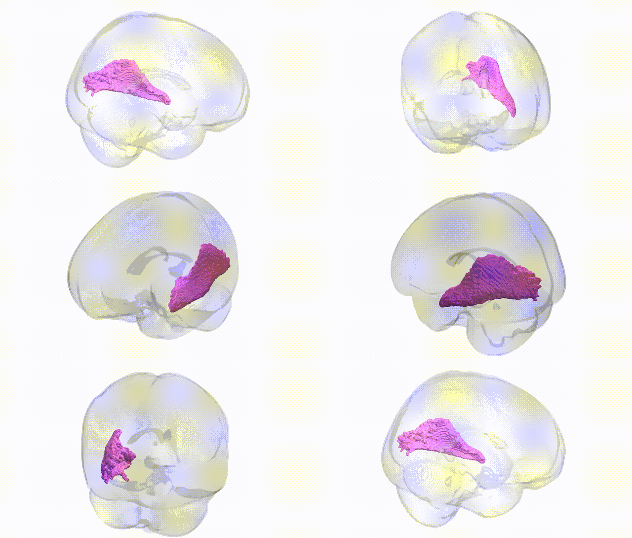
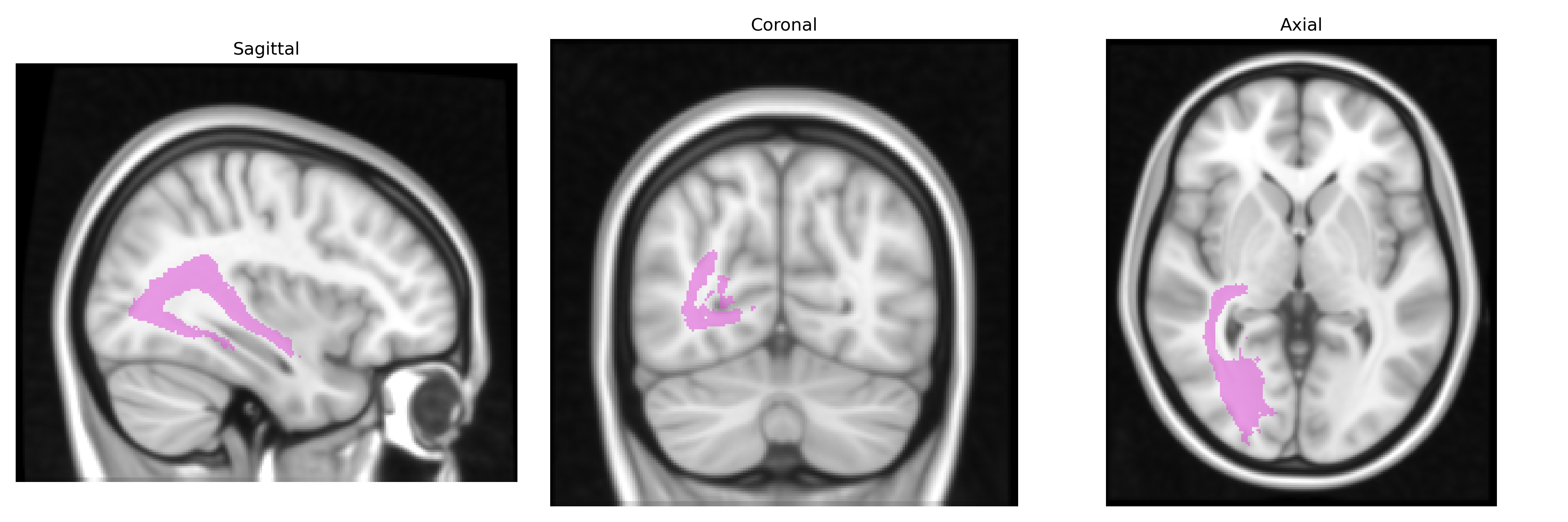
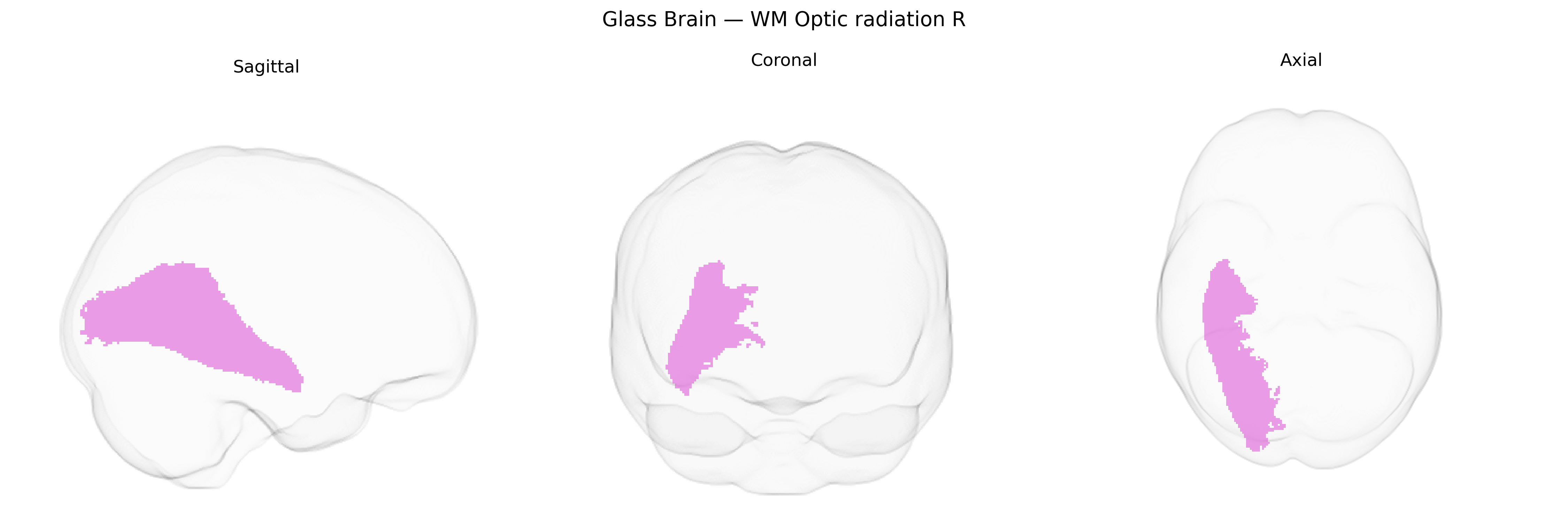
The bilateral WM Optic radiation R from the Juelich maxprob thr25 1mm Atlas corresponds to the right-sided portion of the optic radiations, a major white matter tract conveying visual information from the lateral geniculate nucleus (LGN) of the thalamus to the primary visual cortex (striate cortex, Brodmann area 17) in the occipital lobe. These fibers traverse the parietal and temporal lobes, including Meyer’s loop, and are topographically organized to maintain retinotopic mapping of the visual field. Functionally, this tract is essential for conscious visual perception, transmitting signals related to luminance, contrast, motion, and form, and lesions in this region can result in characteristic visual field defects such as contralateral homonymous hemianopia or quadrantanopia. No direct Wikipedia article exists for this specific atlas-defined region; a closely related structure is the Optic radiation.
The bilateral white matter (WM) of the optic radiation, as defined in the Juelich maxprob thr25 1 mm atlas, has been implicated in several genetic and genome-wide association studies that link its microstructural properties (e.g., fractional anisotropy, mean diffusivity) to neurodevelopmental, neurodegenerative, and psychiatric phenotypes. Large imaging-genetics consortia such as ENIGMA and UK Biobank have reported heritable variation in optic radiation integrity, with common variants in genes involved in axonal guidance, myelination, and synaptic plasticity (for example, NTRK1/2, CNTNAP2, MAG, and other oligodendrocyte-related genes) contributing to individual differences, although single-locus effects are generally small and often sub-threshold after strict correction. GWAS of white-matter tracts and diffusion tensor imaging measures have found that polygenic risk scores for schizophrenia, bipolar disorder, and major depression are associated with reduced integrity of the optic radiation, consistent with broader fronto-temporo-occipital dysconnectivity in these disorders; similar associations have been described for autism spectrum disorder and attention-deficit/hyperactivity disorder at the level of polygenic burden rather than specific loci. In neurodegenerative conditions, variants in genes implicated in Alzheimer’s disease (e.g., APOE) and multiple sclerosis susceptibility loci show associations with optic radiation microstructure, reflecting vulnerability of visual pathways to demyelination and neurodegeneration. GWAS of visual function and visual acuity, as well as of individual differences in brain structural connectivity, also point indirectly to genetic influences on the optic radiation, but no single gene or variant has been established as specifically and uniquely associated with this tract independent of broader white-matter and occipital/temporal connectivity networks.
Overview generated by GPT-4o (2026).
Region ID: 108
Hemisphere: bilateral
Atlas: Juelich maxprob thr25 1mm
Full Quality Version: Download MP4
Full Quality Version: Download MP4
Wali Sidiqyar*, Gaurav Rudravaram*, Elyssa M. McMaster, Trent M. Schwartz, Adam M. Saunders, Kurt G. Schilling, Bennett A. Landman "Introducing SPINS: A Shared Public Visualization Library of Neuroanatomical Structures." Medical Imaging with Deep Learning- short paper
This resource is licensed under CC0 1.0 Universal (Public Domain).