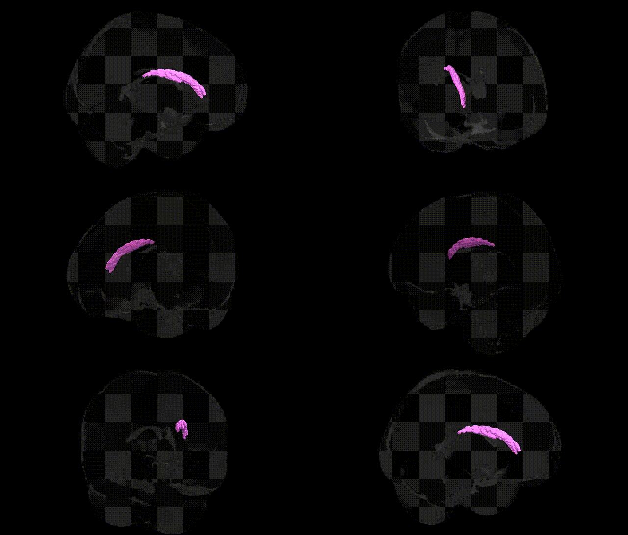
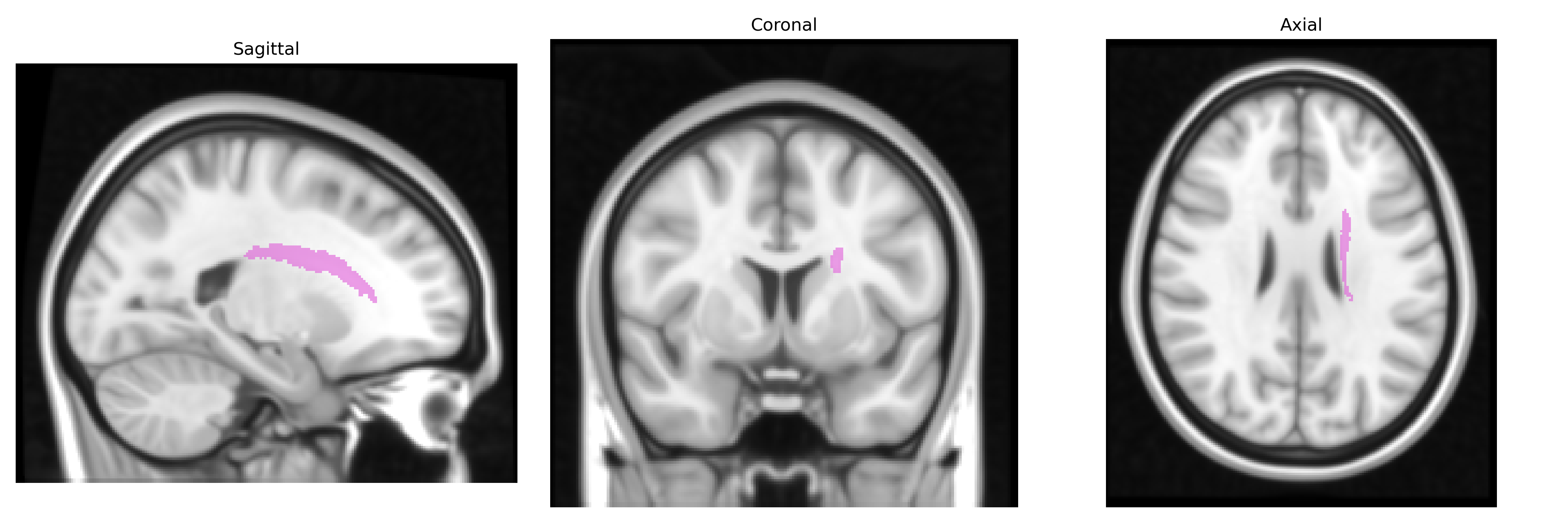
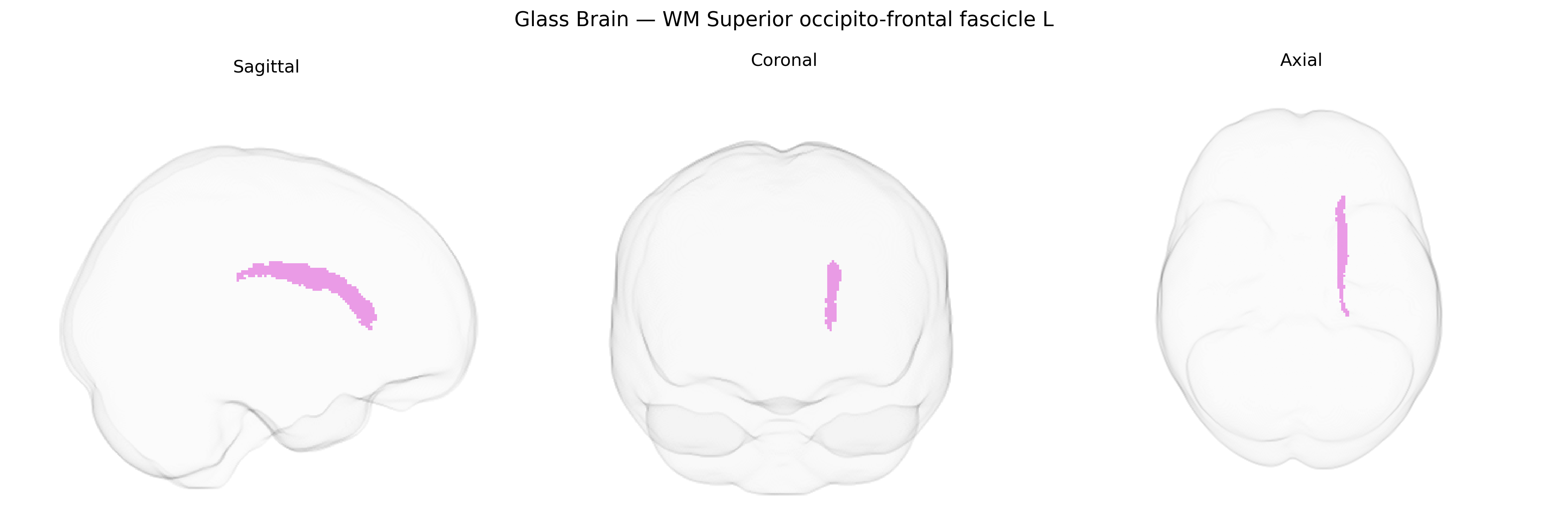
The bilateral WM Superior occipito-frontal fascicle L, as defined in the Juelich maxprob thr25 1mm atlas, corresponds to the left superior fronto-occipital fasciculus, a major long-range association white matter tract that links dorsal and lateral frontal cortices with parietal, occipital, and portions of temporal cortices. This fasciculus runs within the deep white matter lateral to the lateral ventricles, passing through or near the centrum semiovale, and plays a key role in integrating visuospatial, attentional, and higher-order cognitive processes by facilitating communication between posterior sensory regions and anterior executive areas. It is part of the broader fronto-occipital connectivity system and may be implicated in functions such as visuospatial working memory, visual attention, and aspects of language and executive control. There is no direct link for this specific atlas-defined region; a related structure is the Fronto-occipital fasciculus.
The bilateral superior occipito-frontal fascicle (often overlapping with what diffusion MRI studies term the superior longitudinal fasciculus and adjacent fronto-occipital association pathways) has been implicated in several imaging-genetics and GWAS findings, though usually under broader white-matter or tract-based phenotypes rather than the specific Juelich WM Superior occipito-frontal fascicle L label. Large-scale diffusion MRI GWAS (e.g., UK Biobank–based studies by the ENIGMA and related consortia) have reported that fractional anisotropy, mean diffusivity, and other microstructural measures in fronto-occipital and frontoparietal association tracts are heritable and associated with variants near genes involved in axon guidance, myelination, and neurodevelopment (such as genes in the neuregulin/ERBB pathway, oligodendrocyte-related genes, and loci near NCAM1, CNTN4, and other cell-adhesion molecules). Polygenic risk for schizophrenia, bipolar disorder, and major depression has been linked to altered microstructure in superior fronto-occipital and longitudinal fasciculi, suggesting shared genetic architecture between psychiatric risk variants and white-matter integrity; similar associations have been reported for attention-deficit/hyperactivity disorder and autism spectrum traits, where risk loci affecting neurodevelopmental pathways show downstream effects on association-fiber organization. GWAS of general cognitive ability and educational attainment have also identified overlapping genetic influences on frontoparietal and occipito-frontal white-matter integrity, indicating that common variants contributing to higher cognitive performance partly act via microstructural properties of these long-range tracts. However, no single gene or variant is uniquely or specifically associated with the Juelich-defined “WM Superior occipito-frontal fascicle L” region; rather, the region lies within a network of association fibers whose structure reflects polygenic influences on neurodevelopment, myelination, and susceptibility to psychiatric and cognitive phenotypes.
Overview generated by GPT-4o (2026).
Region ID: 113
Hemisphere: bilateral
Atlas: Juelich maxprob thr25 1mm
Full Quality Version: Download MP4
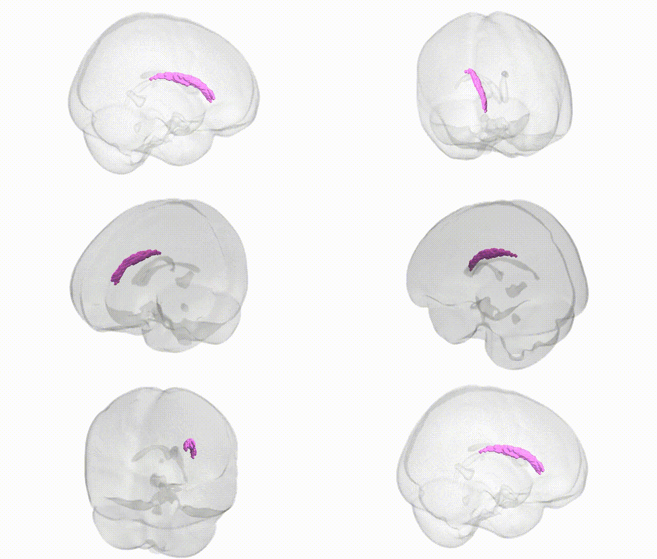
Full Quality Version: Download MP4
Wali Sidiqyar*, Gaurav Rudravaram*, Elyssa M. McMaster, Trent M. Schwartz, Adam M. Saunders, Kurt G. Schilling, Bennett A. Landman "Introducing SPINS: A Shared Public Visualization Library of Neuroanatomical Structures." Medical Imaging with Deep Learning- short paper
This resource is licensed under CC0 1.0 Universal (Public Domain).