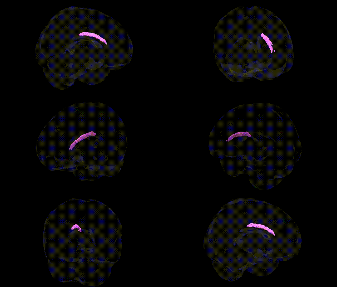
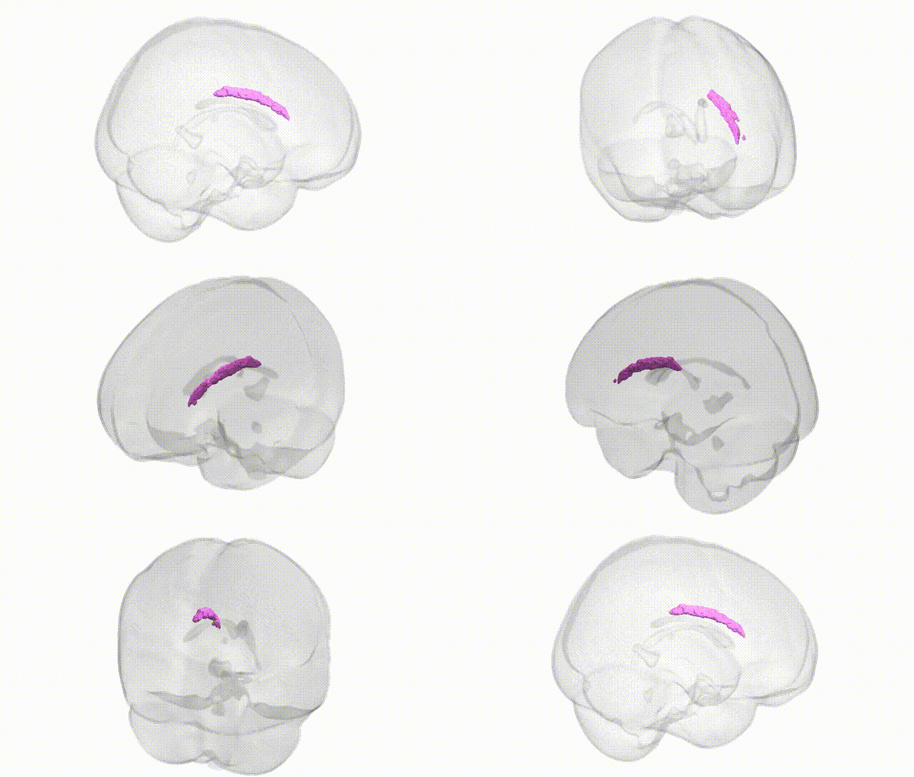
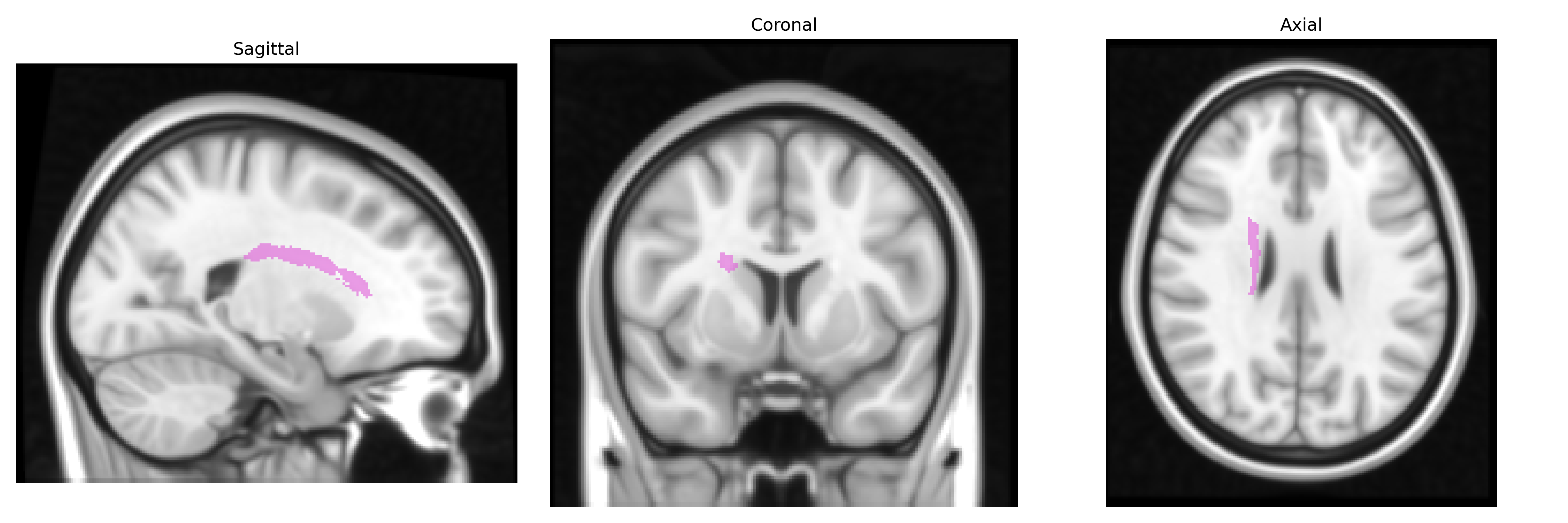
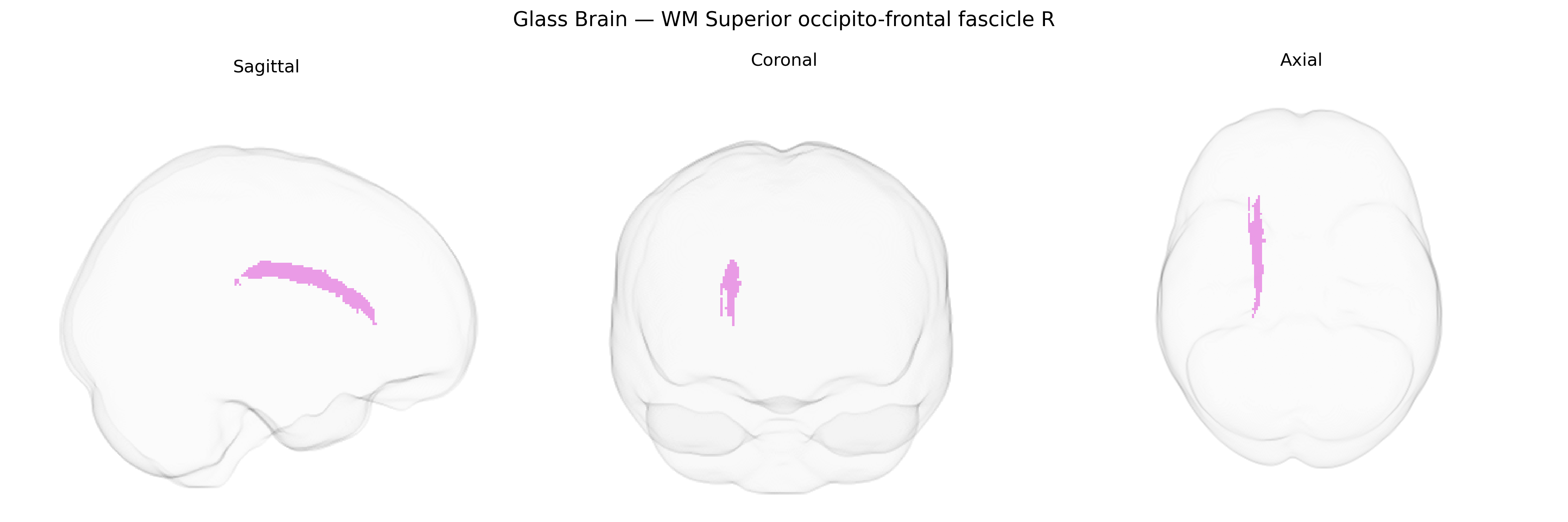
The bilateral WM Superior occipito-frontal fascicle R, as defined in the Juelich maxprob thr25 1mm atlas, corresponds to right-hemisphere long-range association white matter fibers linking posterior occipital and adjacent parietal regions with more anterior frontal cortical areas. This tract supports integration of visual and visuo-spatial information with higher-order executive, planning, and attentional processes by conveying signals between dorsal visual stream regions and frontal control networks. Anatomically, it courses deep to the cortical mantle, traversing the superior part of the hemispheric white matter, and interdigitates with neighboring association bundles such as components of the superior longitudinal fasciculus and other fronto-parietal pathways. There is no direct link for this specific tract; a related structure is the broader Association fiber.
The bilateral white matter superior occipito‑frontal fascicle (often overlapping with or reported as the superior longitudinal fasciculus and adjacent fronto‑occipital tracts in imaging genetics studies) has been implicated in several GWAS of diffusion MRI measures, where common variants in genes involved in axon guidance, myelination, and synaptic plasticity (e.g., CNTNAP2, NTRK1/2, NEUREXIN family, and myelin‑related loci near MAG and MBP) have shown associations with fractional anisotropy or mean diffusivity in fronto‑parietal/occipito‑frontal pathways. Large consortia such as ENIGMA and UK Biobank have identified polygenic influences on microstructure in this tract that overlap with genetic risk for neurodevelopmental and psychiatric conditions, including schizophrenia, bipolar disorder, major depression, ADHD, and autism spectrum disorder, where risk alleles and polygenic risk scores correlate with altered integrity of fronto‑parietal and occipito‑frontal fibers. This region’s microstructure has also been linked in imaging‑genetic work to cognitive traits such as general intelligence, processing speed, working memory, and educational attainment, with GWAS indicating shared genetic architecture between white matter metrics in these tracts and cognitive performance. Additionally, loci associated with neurodegenerative and vascular risk (e.g., APOE, and variants influencing blood pressure and small‑vessel disease) have been related to microstructural changes in long association fibers that include the superior occipito‑frontal fascicle, supporting a genetic contribution to vulnerability for age‑related cognitive decline and white matter lesions in this pathway.
Overview generated by GPT-4o (2026).
Region ID: 112
Hemisphere: bilateral
Atlas: Juelich maxprob thr25 1mm
Full Quality Version: Download MP4
Full Quality Version: Download MP4
Wali Sidiqyar*, Gaurav Rudravaram*, Elyssa M. McMaster, Trent M. Schwartz, Adam M. Saunders, Kurt G. Schilling, Bennett A. Landman "Introducing SPINS: A Shared Public Visualization Library of Neuroanatomical Structures." Medical Imaging with Deep Learning- short paper
This resource is licensed under CC0 1.0 Universal (Public Domain).