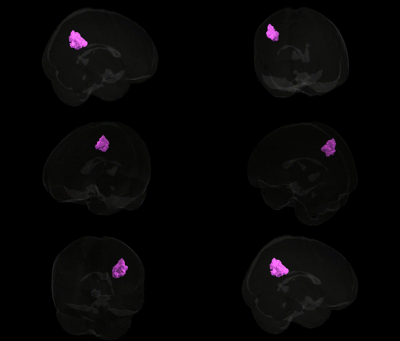
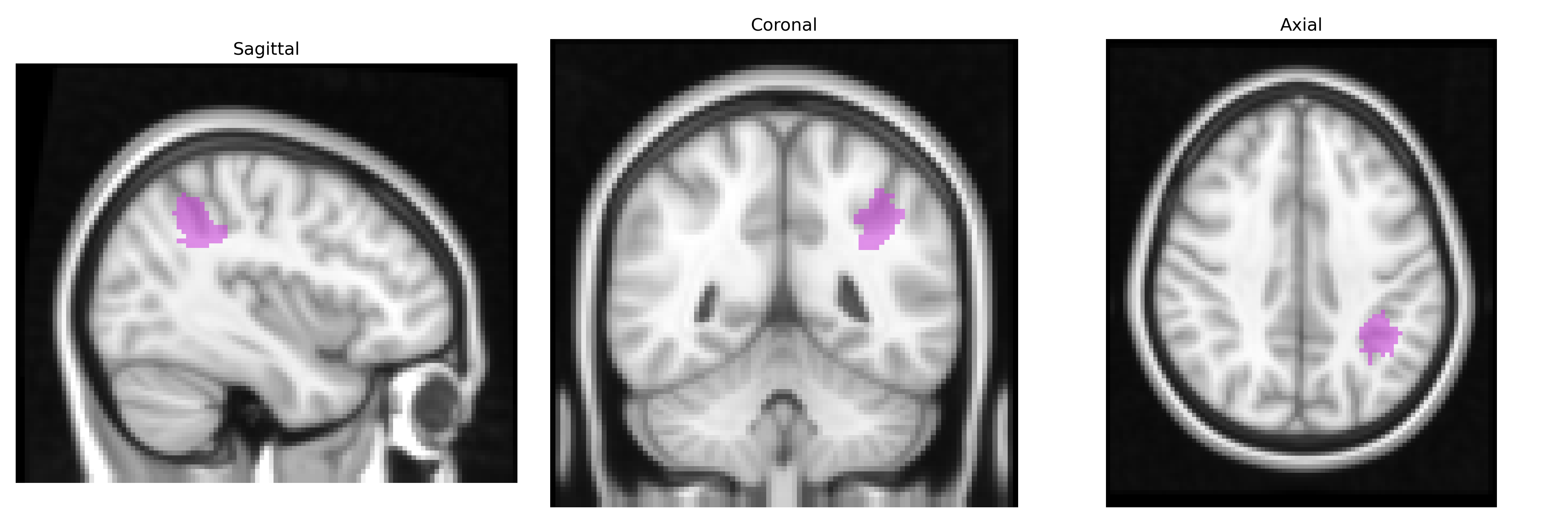
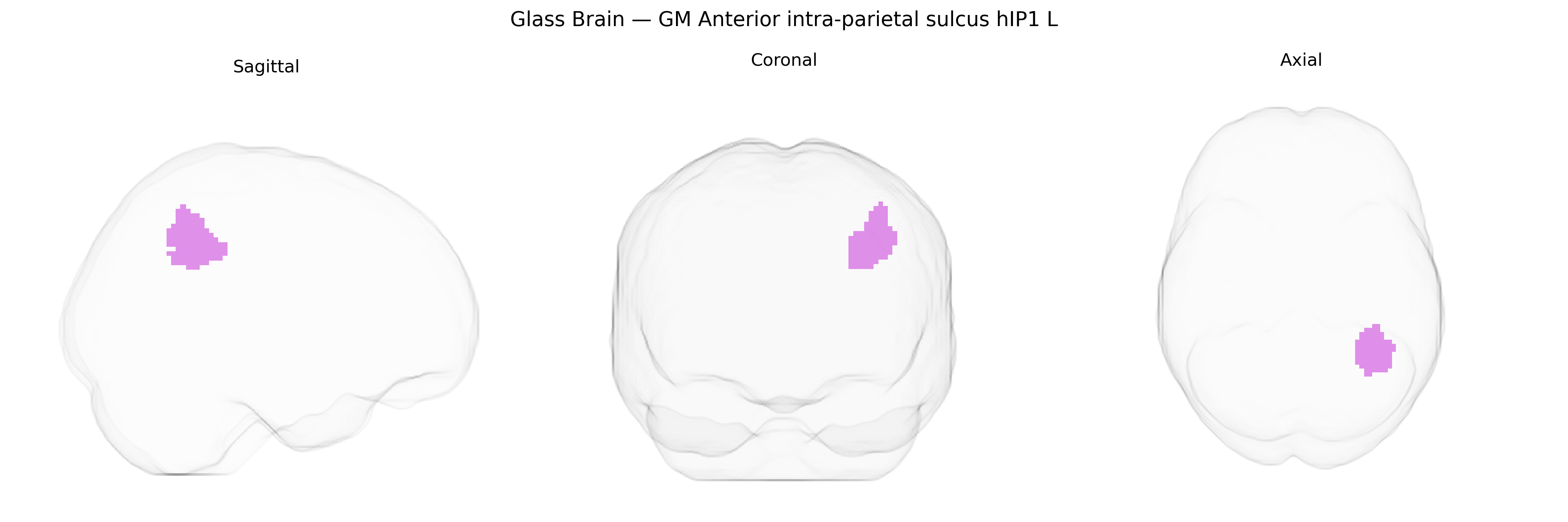
The bilateral GM Anterior intra-parietal sulcus hIP1 L region, as defined in the Juelich maxprob thr25 2 mm atlas, corresponds to a cytoarchitectonically defined subregion of the human intraparietal sulcus located in the inferior parietal lobule, associated with visuomotor integration, object-directed actions, and aspects of numerosity and attention processing. This area is part of the broader intraparietal sulcus complex and is thought to contribute to transforming visual information into coordinated hand and eye movements, including grasping and manipulation behaviors, as well as higher-order perceptual and cognitive functions involving spatial representation. There is no direct Wikipedia article for hIP1; a related structure within which it is embedded is the Intraparietal sulcus.
The anterior intraparietal sulcus (hIP1) is a parietal association region implicated in visuomotor integration, attention, and higher-order cognitive control, and GWAS-based imaging genetics studies have linked structural and functional variation in this area to a range of polygenic influences rather than single-gene effects. Large-scale neuroimaging GWAS from consortia such as ENIGMA and UK Biobank have associated parietal cortical thickness, surface area, and gyrification—often overlapping with the intraparietal sulcus—with common variants in loci including KIAA0586, MEF2C, DAAM1, and genes involved in neuronal development, synaptic plasticity, and cytoskeletal organization; these variants collectively contribute to general cognitive ability, educational attainment, and intelligence, traits that show robust parietal cortex associations. Moreover, polygenic risk scores for neurodevelopmental and psychiatric conditions such as schizophrenia, ADHD, autism spectrum disorder, and major depressive disorder have been linked to altered parietal morphology and connectivity patterns encompassing the intraparietal sulcus in GWAS-informed imaging studies, although these effects are typically modest and distributed across widespread cortical networks. Specific task-based and resting-state fMRI GWAS highlight that variants associated with attention, working memory, and visuospatial performance often show functional correlates in parietal regions including hIP1, but at present there are no well-established, hIP1-specific single-gene associations; rather, bilateral GM hIP1 emerges as a node within polygenically influenced frontoparietal circuits mediating cognition and susceptibility to multiple complex brain disorders.
Overview generated by GPT-4o (2026).
Region ID: 1
Hemisphere: bilateral
Atlas: Juelich maxprob thr25 2mm
Full Quality Version: Download MP4
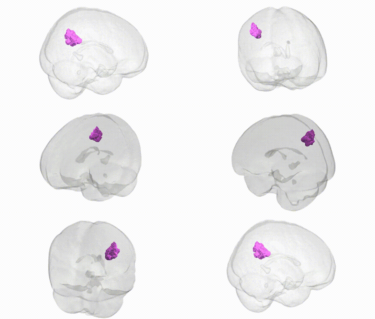
Full Quality Version: Download MP4
Wali Sidiqyar*, Gaurav Rudravaram*, Elyssa M. McMaster, Trent M. Schwartz, Adam M. Saunders, Kurt G. Schilling, Bennett A. Landman "Introducing SPINS: A Shared Public Visualization Library of Neuroanatomical Structures." Medical Imaging with Deep Learning- short paper
This resource is licensed under CC0 1.0 Universal (Public Domain).