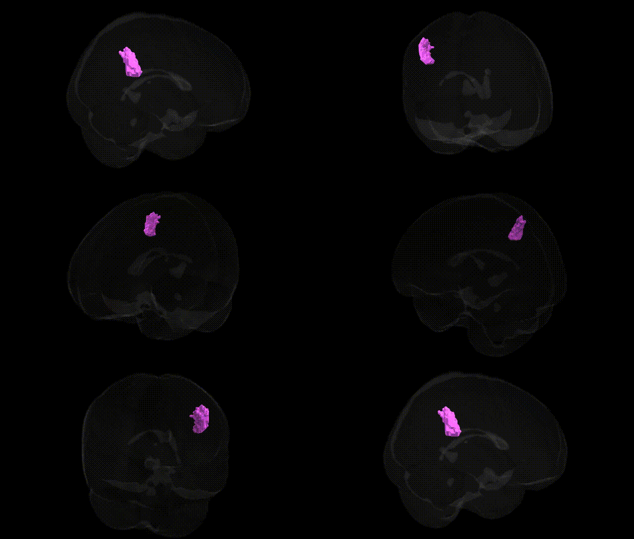
The bilateral GM Anterior intra-parietal sulcus hIP2 L region, as defined in the Juelich maxprob thr25 2mm Atlas, corresponds to a cytoarchitectonically and functionally characterized subregion within the human intraparietal sulcus of the posterior parietal cortex. hIP2 is implicated in visuospatial attention, eye–hand coordination, and sensorimotor integration, particularly in transforming visual information into motor plans for reaching and grasping. It participates in dorsal stream processing and contributes to spatial working memory, object-directed action planning, and integration of visual and proprioceptive signals. While there is no direct Wikipedia article for hIP2, it is part of the broader intraparietal sulcus complex: Intraparietal sulcus.
The bilateral GM Anterior intra-parietal sulcus hIP2 L region from the Juelich maxprob thr25 2mm atlas lies within the anterior intraparietal sulcus (AIP) / inferior parietal lobule, a multimodal hub implicated in visuomotor integration, attention, and higher-order cognition, and genetic associations identified for parietal and intraparietal cortex generally extend to this area. Large-scale GWAS of cortical surface area and thickness (e.g., ENIGMA, UK Biobank) have linked common variants near genes involved in neurodevelopment, synaptic function, and axon guidance—such as MAPT, MIR137, MEF2C, and multiple loci on chromosomes 3p, 6p, 17q, and 19p—with structural variation in parietal and inferior parietal regions that encompass hIP2. Parietal association cortex, including the intraparietal sulcus, frequently appears as part of networks (frontoparietal, dorsal attention, and default mode) whose connectivity and morphology show genetic correlations with cognitive ability, educational attainment, and neuropsychiatric risk (notably schizophrenia, major depression, attention-deficit/hyperactivity disorder, and autism spectrum disorder), often mediated by polygenic scores for these traits. Imaging–genetics studies also implicate this region in genetic risk for Alzheimer’s disease and other dementias, where APOE ε4 and loci near CLU and PICALM show associations with parietal atrophy or altered connectivity in frontoparietal and default mode networks. Although no single gene has been uniquely tied to hIP2 in isolation, convergent GWAS and imaging-genetics findings indicate that genetic variation influencing synaptic plasticity, neurodevelopmental patterning, and neurodegenerative vulnerability contributes to individual differences in structure and function of the anterior intraparietal sulcus, with downstream links to cognition, psychiatric illness, and neurodegenerative disorders.
Overview generated by GPT-4o (2026).
Region ID: 3
Hemisphere: bilateral
Atlas: Juelich maxprob thr25 2mm
Full Quality Version: Download MP4
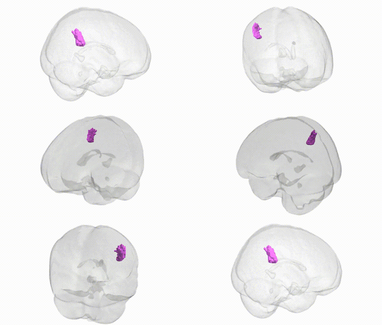
Full Quality Version: Download MP4
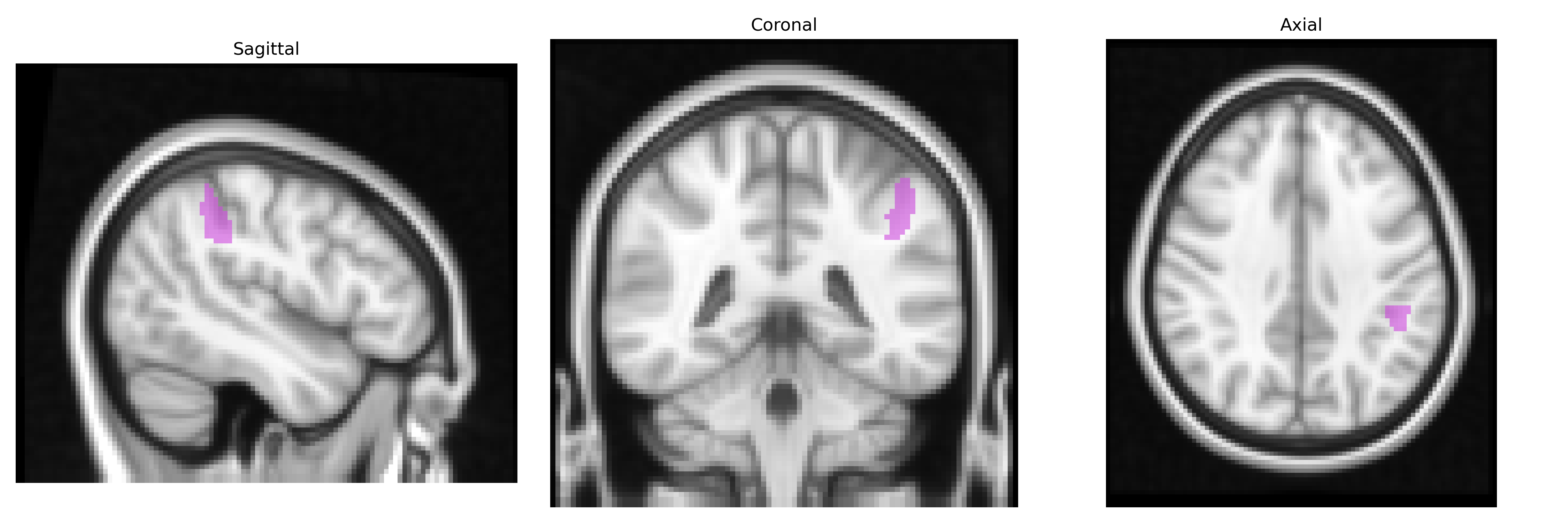
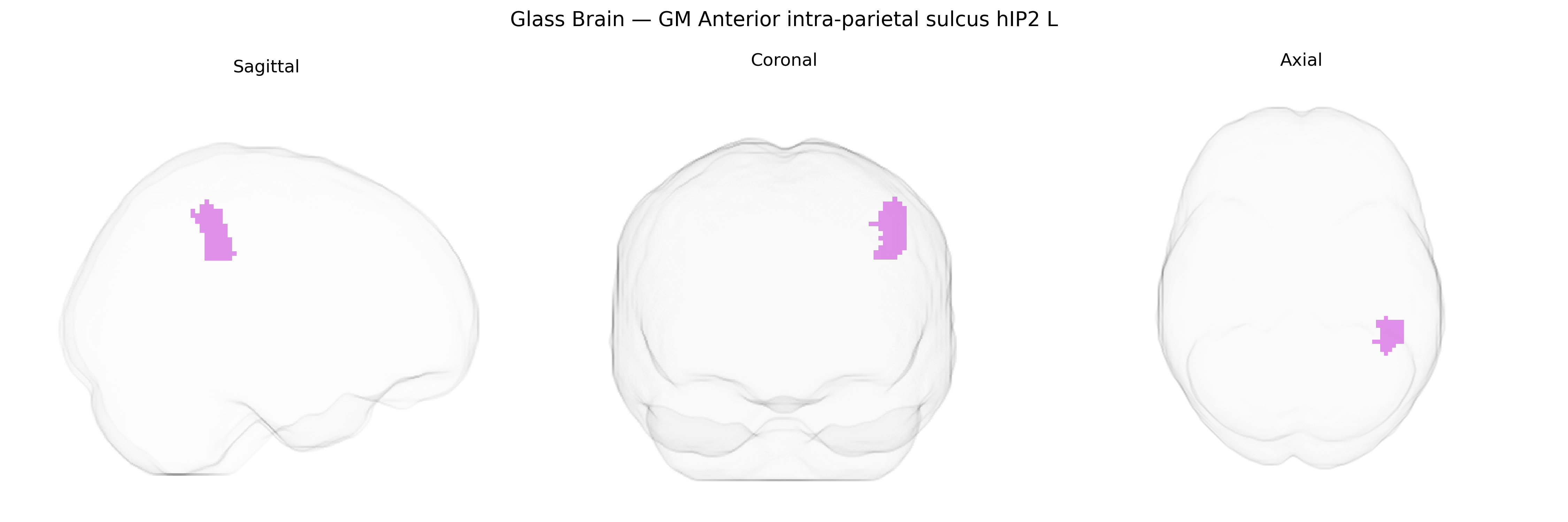
Wali Sidiqyar*, Gaurav Rudravaram*, Elyssa M. McMaster, Trent M. Schwartz, Adam M. Saunders, Kurt G. Schilling, Bennett A. Landman "Introducing SPINS: A Shared Public Visualization Library of Neuroanatomical Structures." Medical Imaging with Deep Learning- short paper
This resource is licensed under CC0 1.0 Universal (Public Domain).