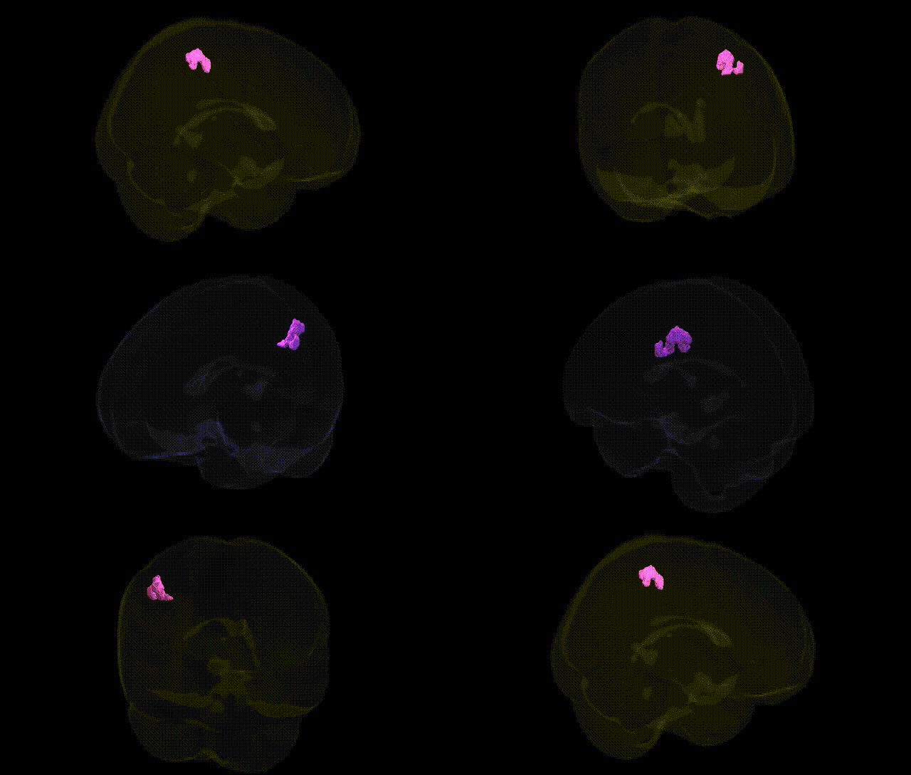
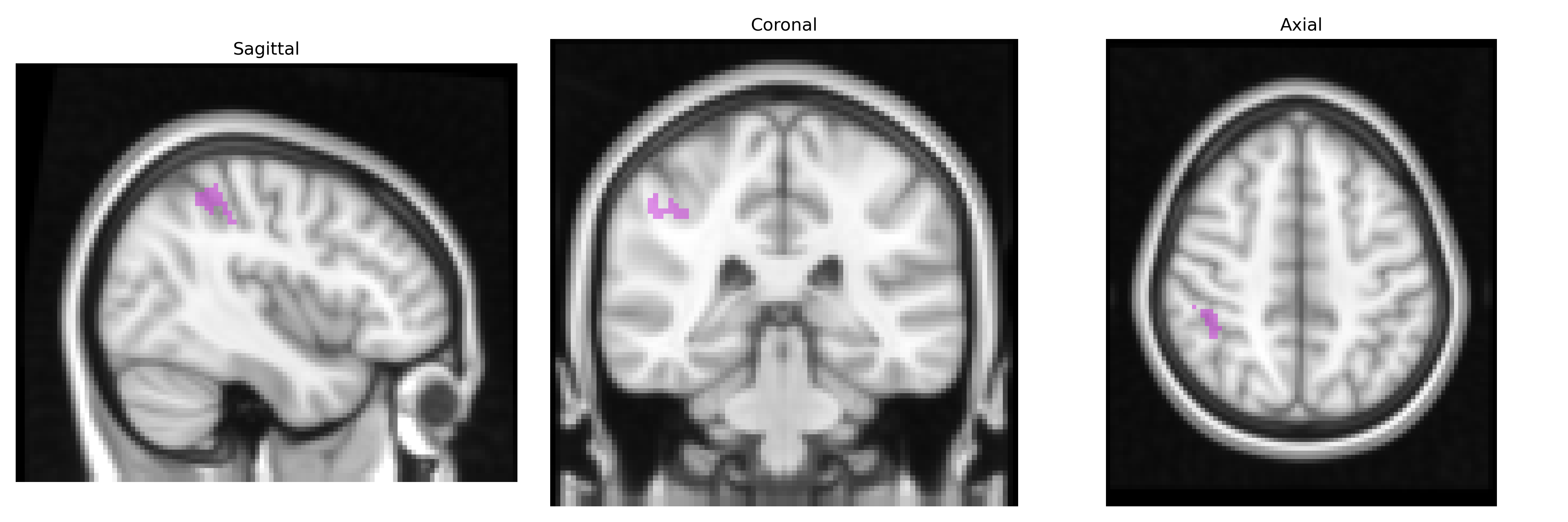
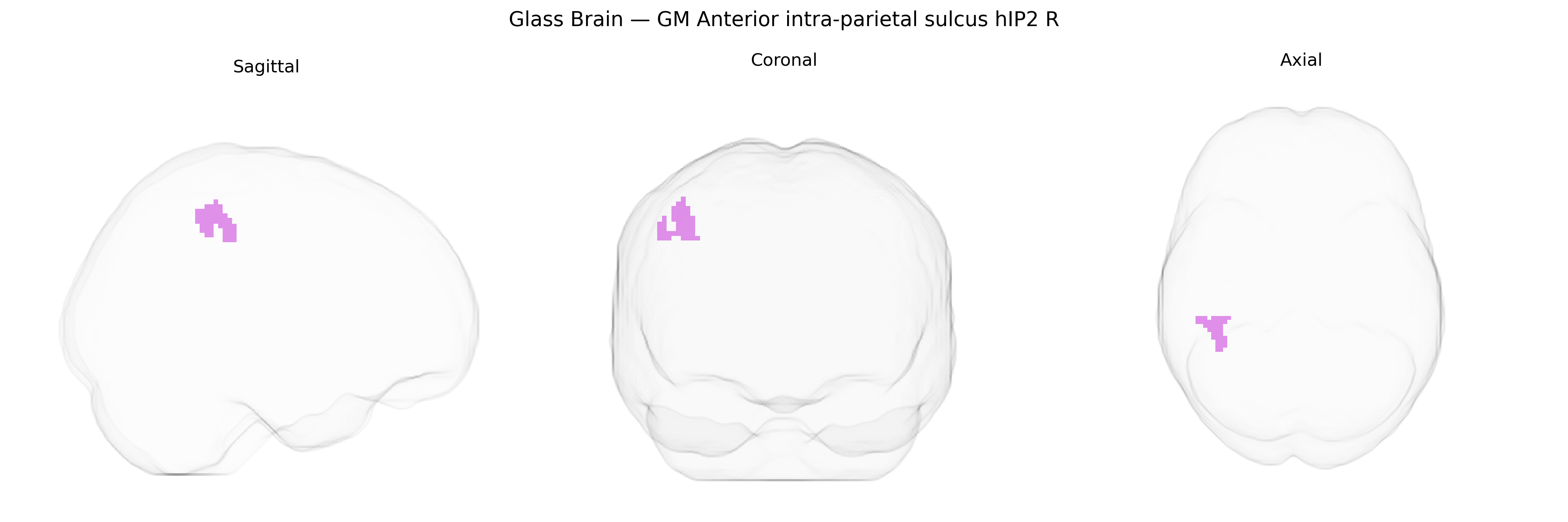
The bilateral GM Anterior intra-parietal sulcus hIP2 R region, as defined in the Juelich maxprob thr25 2mm Atlas, corresponds to a cytoarchitectonically and functionally characterized subregion of the human intraparietal sulcus within the posterior parietal cortex, predominantly in the right hemisphere. It is implicated in visuomotor integration, object-directed actions, and higher-order spatial processing, contributing to functions such as grasp planning, hand–eye coordination, and the transformation of visual information into motor commands. This region forms part of the broader intraparietal sulcus network that interfaces with dorsal visual and premotor areas, supporting sensorimotor transformations necessary for goal-directed behavior. There is no direct link for hIP2; a related area within the same sulcus is described here: Intraparietal sulcus.
The bilateral GM anterior intra-parietal sulcus hIP2 (R) region, as defined in the Juelich maxprob thr25 2mm atlas, lies within the broader intraparietal sulcus (IPS), where genetic associations have primarily been inferred from GWAS of cognitive, perceptual, and neuropsychiatric traits rather than from region-specific SNP analyses. Variants in genes influencing synaptic plasticity, cortical development, and parietal lobe morphology (such as BDNF, DISC1, CNTNAP2, and common polygenic influences captured by genome-wide polygenic scores) have been linked to IPS structure and function, including visuospatial attention, numerical processing, and working memory, functions that overlap with the putative role of hIP2. Large-scale imaging genetics studies (e.g., ENIGMA and UK Biobank) have reported heritable variation in parietal cortical thickness and surface area, with multiple loci across the genome contributing to IPS anatomy, although these effects are typically mapped to broader parietal regions rather than to hIP2 specifically. GWAS of mathematical ability and numeracy show associations with parietal activation patterns, including the IPS, and implicate polygenic influences distributed across neurodevelopmental and synaptic genes, while studies of attention-deficit/hyperactivity disorder, schizophrenia, and autism spectrum disorder have identified parietal and IPS alterations that are partly explained by known risk variants and polygenic burden, again at a coarse regional level. Task-based fMRI GWAS linking genetic variation to IPS activation during working memory, attention, and decision-making tasks support a distributed, highly polygenic architecture with no single gene uniquely or consistently tied to hIP2, indicating that genetic associations relevant to this region are best understood as part of the broader parietal and frontoparietal network rather than as region-specific findings.
Overview generated by GPT-4o (2026).
Region ID: 4
Hemisphere: bilateral
Atlas: Juelich maxprob thr25 2mm
Full Quality Version: Download MP4
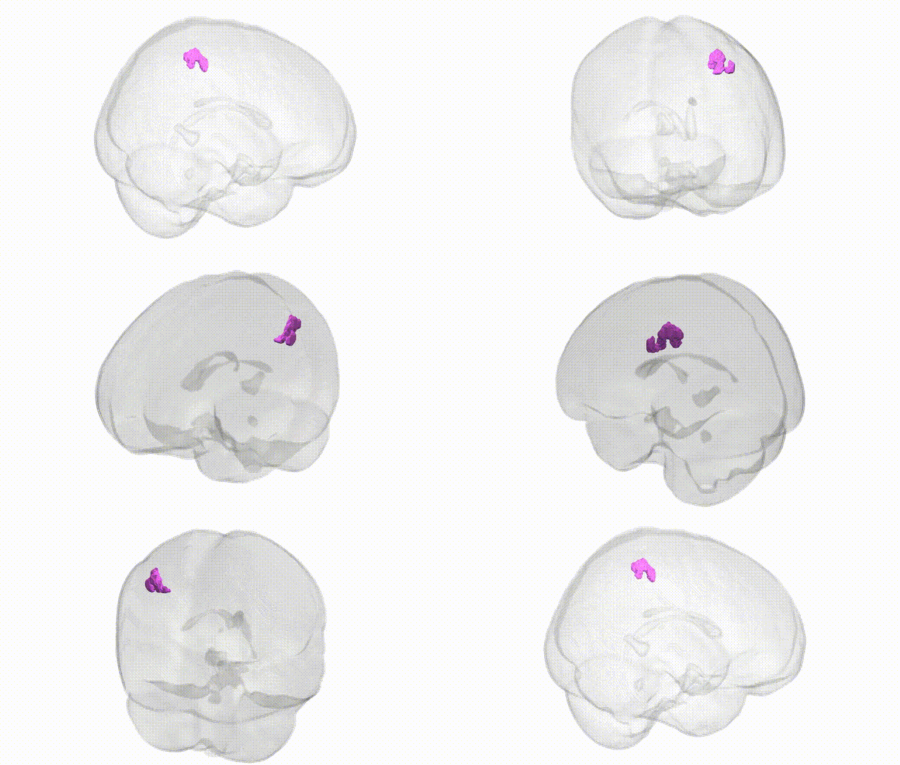
Full Quality Version: Download MP4
Wali Sidiqyar*, Gaurav Rudravaram*, Elyssa M. McMaster, Trent M. Schwartz, Adam M. Saunders, Kurt G. Schilling, Bennett A. Landman "Introducing SPINS: A Shared Public Visualization Library of Neuroanatomical Structures." Medical Imaging with Deep Learning- short paper
This resource is licensed under CC0 1.0 Universal (Public Domain).