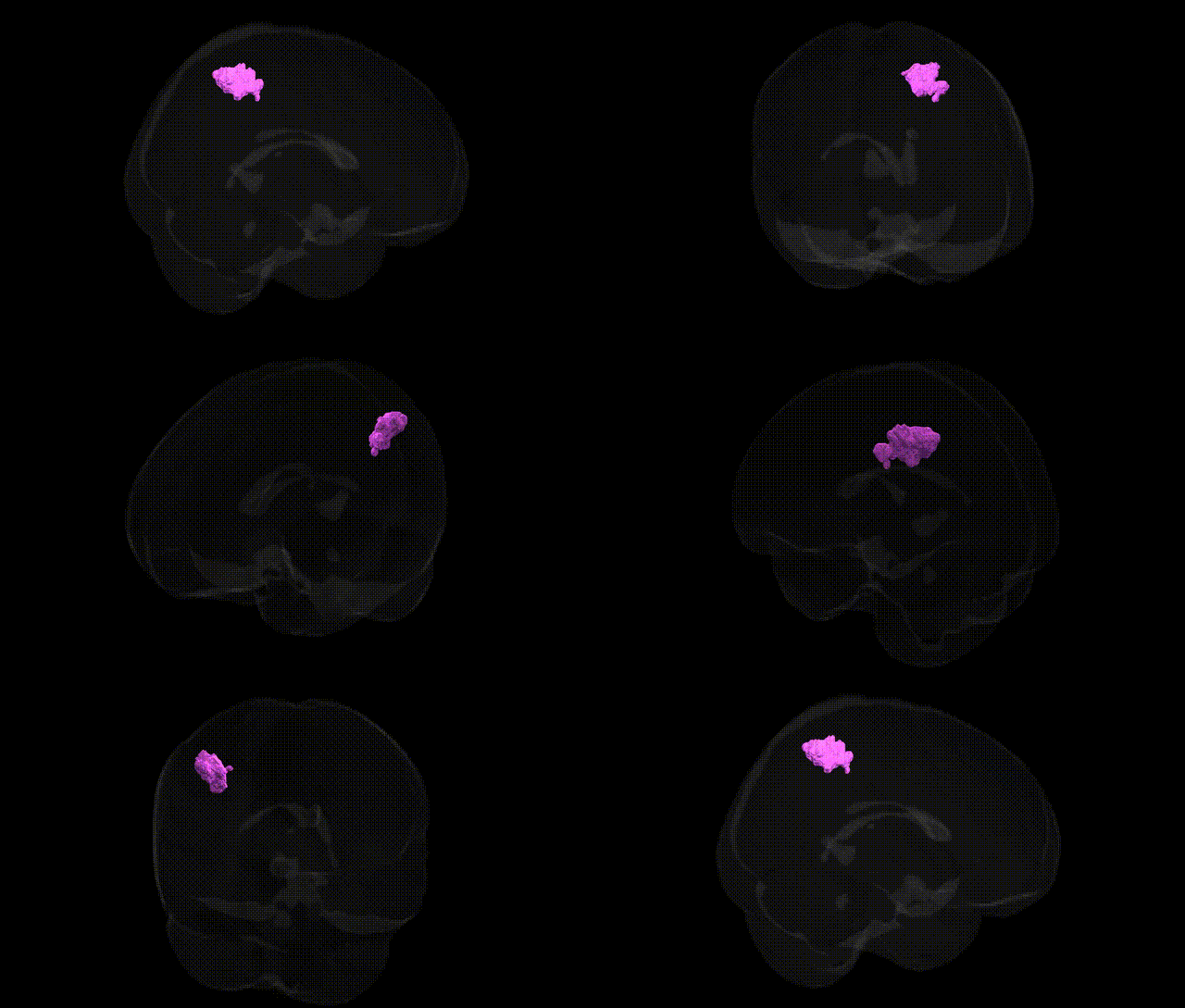
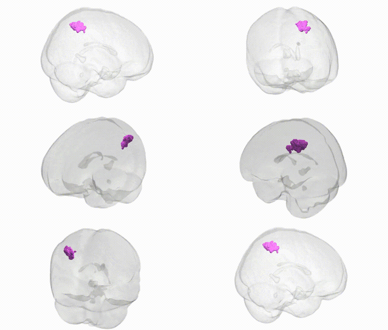
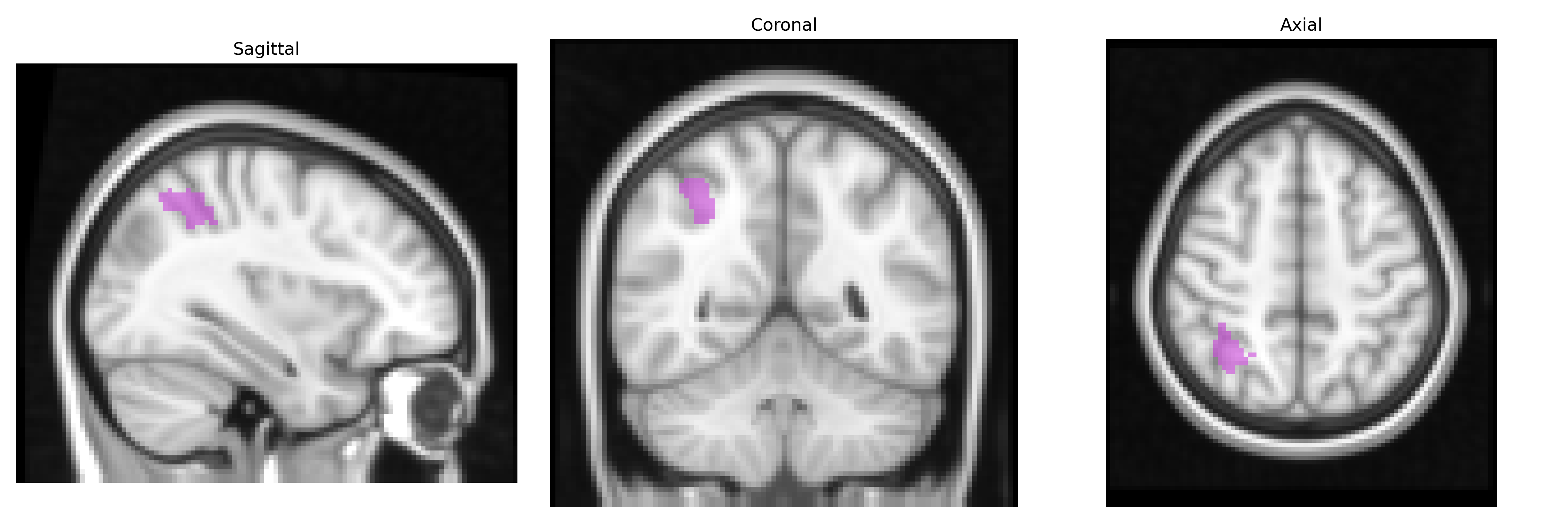
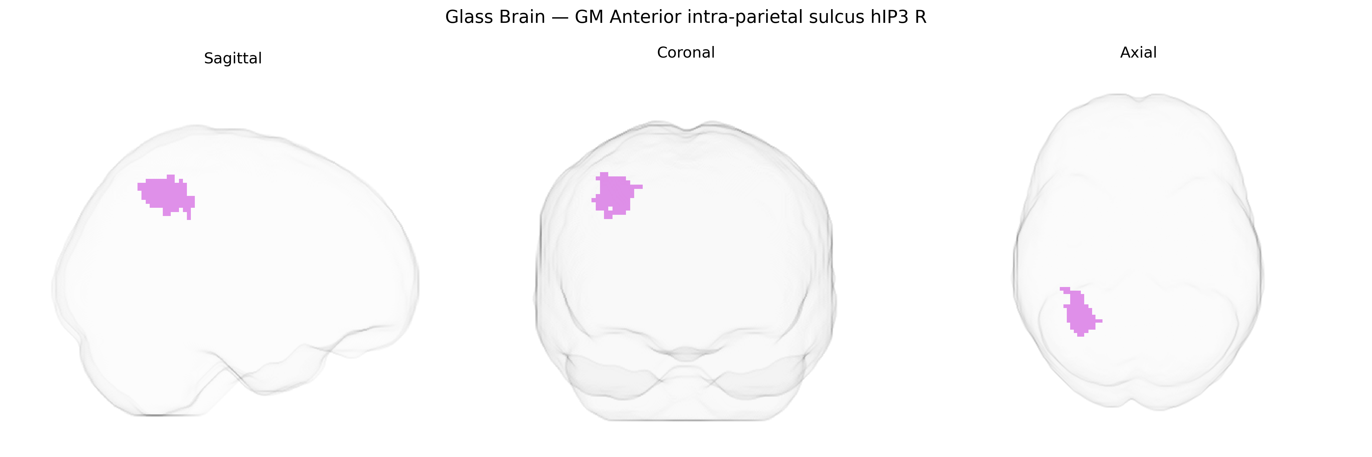
The bilateral GM Anterior intra-parietal sulcus hIP3 R region in the Juelich maxprob thr25 2mm atlas corresponds to a cytoarchitectonically defined subregion of the intraparietal sulcus within the posterior parietal cortex, associated with higher-order visuomotor integration, object-directed actions, and aspects of attention and spatial processing. This area is implicated in transforming visual information into motor commands for grasping and manipulating objects, integrating sensory input with motor plans, and contributing to the representation of object features such as size, shape, and orientation. As a probabilistic map derived from post-mortem cytoarchitecture, hIP3 R reflects interindividual variability in the parietal lobe and is functionally linked to the broader fronto-parietal network involved in perception–action coupling and visuo-spatial cognition. There is no direct Wikipedia article for this specific subregion; a related structure is the broader Intraparietal sulcus.
The bilateral GM Anterior intra-parietal sulcus hIP3 region, as defined in the Juelich maxprob thr25 2mm atlas, lies within the broader intraparietal sulcus (IPS) network implicated in visuospatial attention, eye–hand coordination, and higher-order cognitive functions, and genetic associations for this area derive largely from imaging genetics and GWAS of cortical morphology, functional activation, and related traits rather than region-specific candidate studies. Large-scale GWAS of cortical thickness and surface area (e.g., ENIGMA, UK Biobank) have identified multiple loci—such as variants near RSPO3, TESC, DACT1, HMGA2, and genes influencing Wnt signaling, neurodevelopment, and synaptic function—associated with parietal cortex structure that encompasses the hIP3 field, while task-based fMRI GWAS of attention and working memory have linked polygenic influences on frontoparietal network engagement to genes involved in dopaminergic and glutamatergic signaling. Parietal regions including IPS/hIP3 show heritable variability and appear as mediators in imaging-genetic studies of neurodevelopmental and psychiatric conditions, with schizophrenia, ADHD, and autism polygenic risk scores associated with altered IPS activation and morphology, and Alzheimer’s disease and other dementias showing parietal atrophy patterns that partially overlap hIP3 and correlate with APOE and other risk loci. Additionally, GWAS of cognitive traits and educational attainment indicate that parietal cortical measures, including IPS involvement, partly mediate the effects of polygenic scores for intelligence and learning, suggesting that hIP3 serves as a genetically influenced hub linking common neurodevelopmental pathways to attentional control, visuospatial processing, and higher cognition, though direct GWAS targeting this specific Juelich subregion remain limited and findings are typically reported at the level of broader parietal or frontoparietal territories.
Overview generated by GPT-4o (2026).
Region ID: 6
Hemisphere: bilateral
Atlas: Juelich maxprob thr25 2mm
Full Quality Version: Download MP4
Full Quality Version: Download MP4
Wali Sidiqyar*, Gaurav Rudravaram*, Elyssa M. McMaster, Trent M. Schwartz, Adam M. Saunders, Kurt G. Schilling, Bennett A. Landman "Introducing SPINS: A Shared Public Visualization Library of Neuroanatomical Structures." Medical Imaging with Deep Learning- short paper
This resource is licensed under CC0 1.0 Universal (Public Domain).