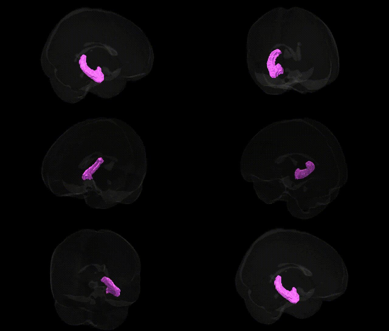
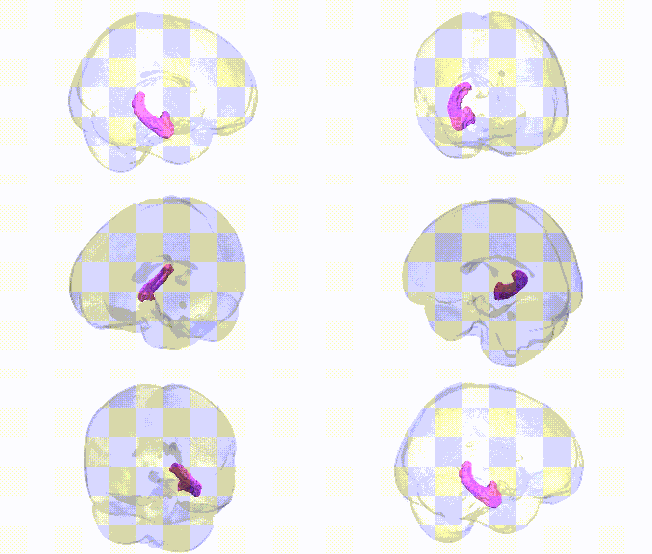
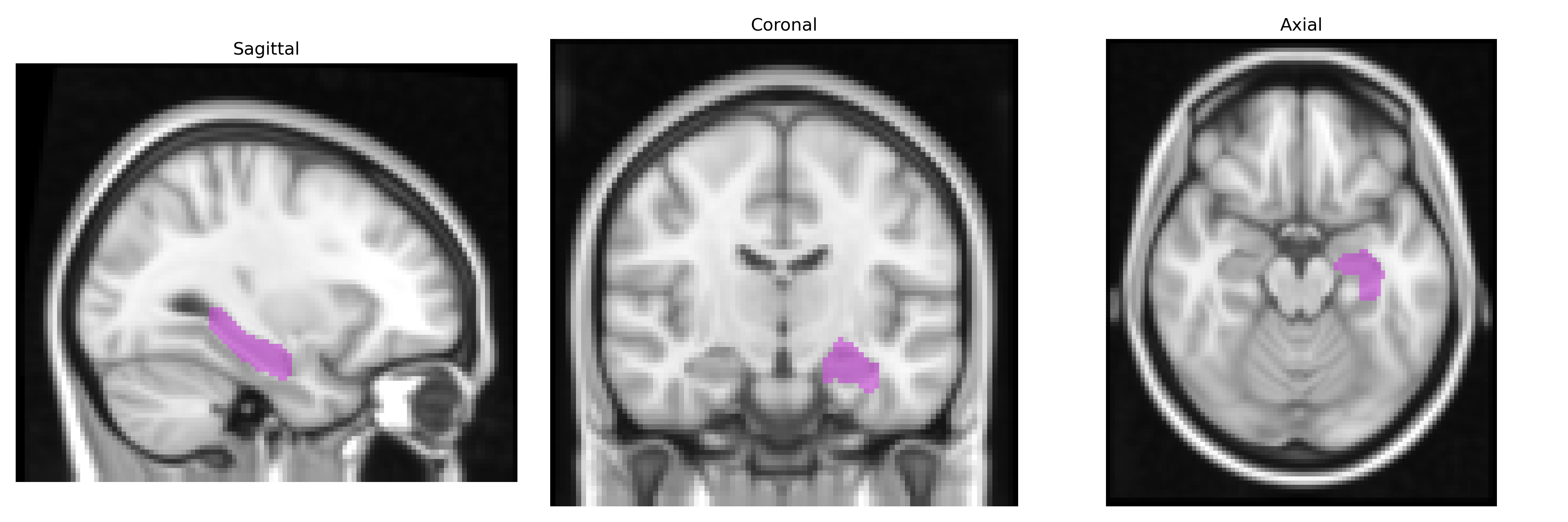
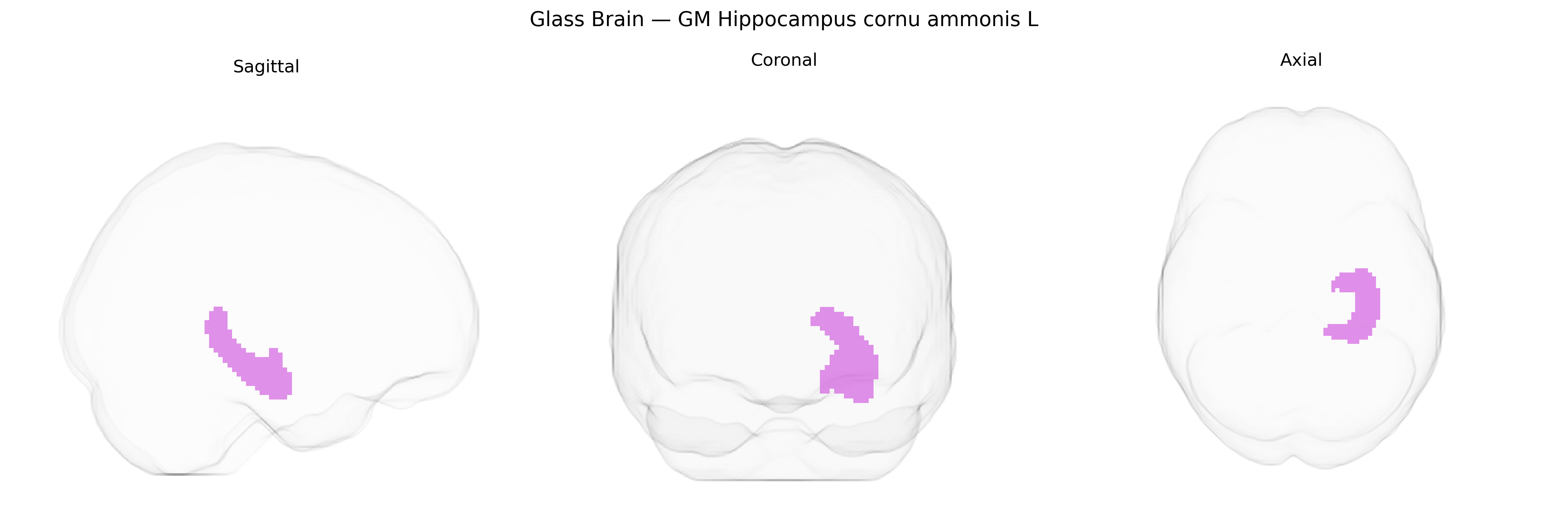
The bilateral GM Hippocampus cornu ammonis L region in the Juelich maxprob thr25 2mm Atlas corresponds to the gray matter of the left cornu ammonis (CA) fields of the hippocampus, a key allocortical structure located in the medial temporal lobe and forming part of the hippocampal formation. It comprises distinct subfields (e.g., CA1–CA4) characterized by layered pyramidal neurons and specialized microcircuitry that support synaptic plasticity and long-term potentiation. Functionally, this region is critically involved in episodic memory encoding and retrieval, spatial navigation, sequence learning, and the consolidation of information from short- to long-term memory, while also contributing to stress regulation via dense glucocorticoid receptor expression. The cornu ammonis is tightly interconnected with the dentate gyrus, subiculum, entorhinal cortex, and broader limbic networks, making it highly susceptible to neurodegenerative, ischemic, and epileptogenic processes. A related reference entry is Hippocampus.
The bilateral gray matter of the hippocampal cornu ammonis (CA) fields, as delineated in the Juelich maxprob thr25 2 mm atlas, has been repeatedly implicated in genetic studies of hippocampal volume, structure, and function, with numerous genome-wide association studies (GWAS) identifying common variants that influence this region. Large neuroimaging genetics consortia (e.g., ENIGMA, UK Biobank–based GWAS) have reported loci in or near genes such as SLC4A10, HMGA2, DPP4, APOE, BDNF, and others as contributors to hippocampal volume and subfield morphology, including CA regions, with several of these variants also associated with cognitive performance, memory, and general intelligence. APOE ε4 and other Alzheimer’s disease–related variants show robust links to reduced hippocampal and CA subfield volumes, accelerated atrophy, and increased risk of late-onset Alzheimer’s disease, while schizophrenia, major depressive disorder, and bipolar disorder GWAS have identified polygenic risk profiles that correlate with reduced hippocampal integrity and specific CA subfield abnormalities. GWAS of PTSD, anxiety traits, and stress reactivity further implicate variants in stress-response and synaptic plasticity genes (e.g., FKBP5, CRHR1, and glutamatergic signaling genes) in altered CA subfield structure and function, paralleling the key role of these fields in fear conditioning and contextual memory. Additionally, genetic influences on hippocampal neurogenesis and synaptic remodeling, including variants in cell adhesion, axon guidance, and neurotrophic pathways, have been associated with resilience or vulnerability to neurodegenerative and psychiatric disorders, underscoring the CA hippocampal subfields as a central convergence point for genetic effects on memory, emotion, and disease risk.
Overview generated by GPT-4o (2026).
Region ID: 17
Hemisphere: bilateral
Atlas: Juelich maxprob thr25 2mm
Full Quality Version: Download MP4
Full Quality Version: Download MP4
Wali Sidiqyar*, Gaurav Rudravaram*, Elyssa M. McMaster, Trent M. Schwartz, Adam M. Saunders, Kurt G. Schilling, Bennett A. Landman "Introducing SPINS: A Shared Public Visualization Library of Neuroanatomical Structures." Medical Imaging with Deep Learning- short paper
This resource is licensed under CC0 1.0 Universal (Public Domain).