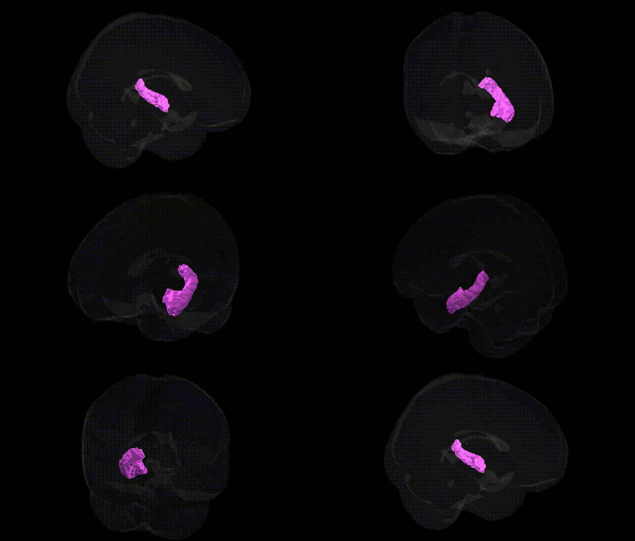
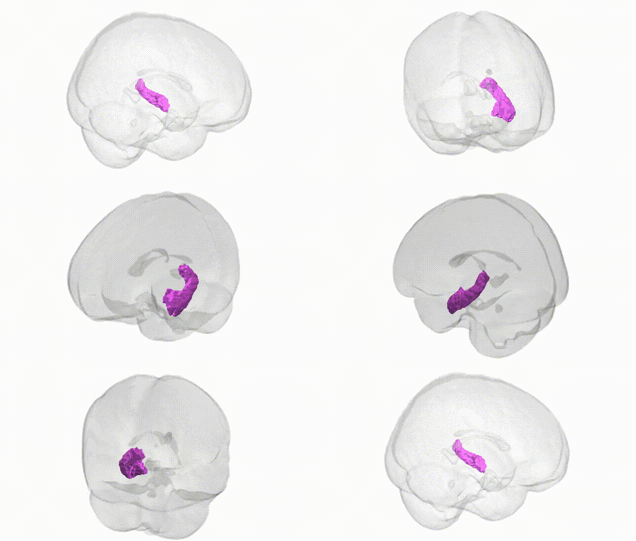
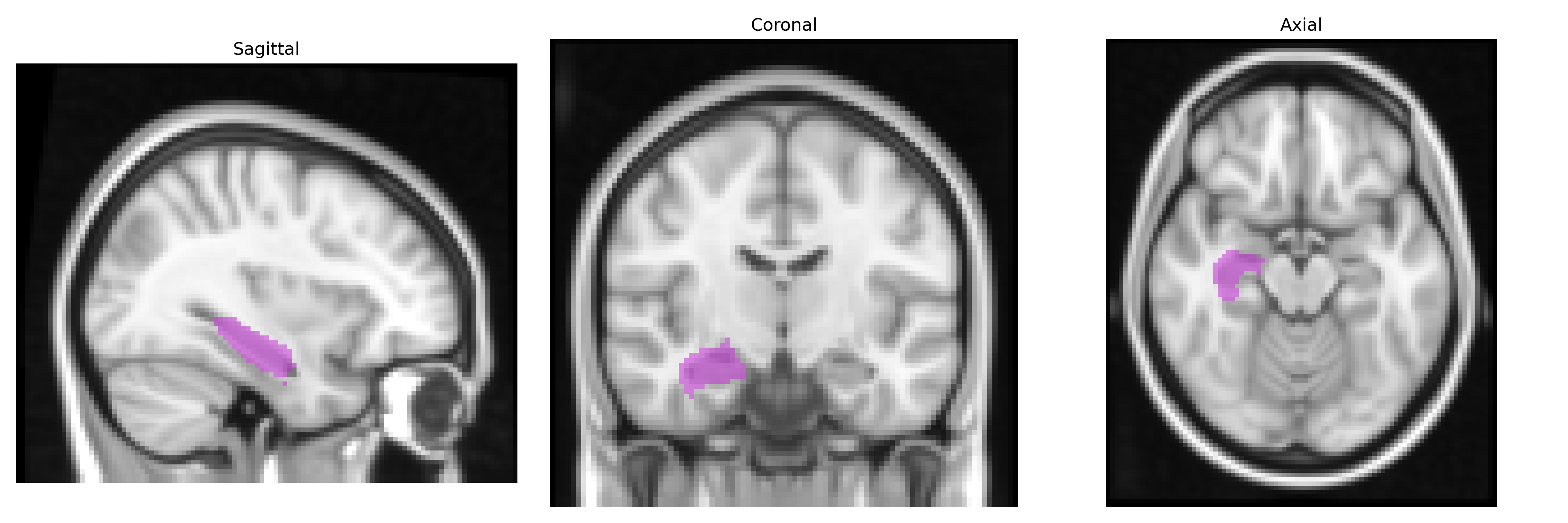
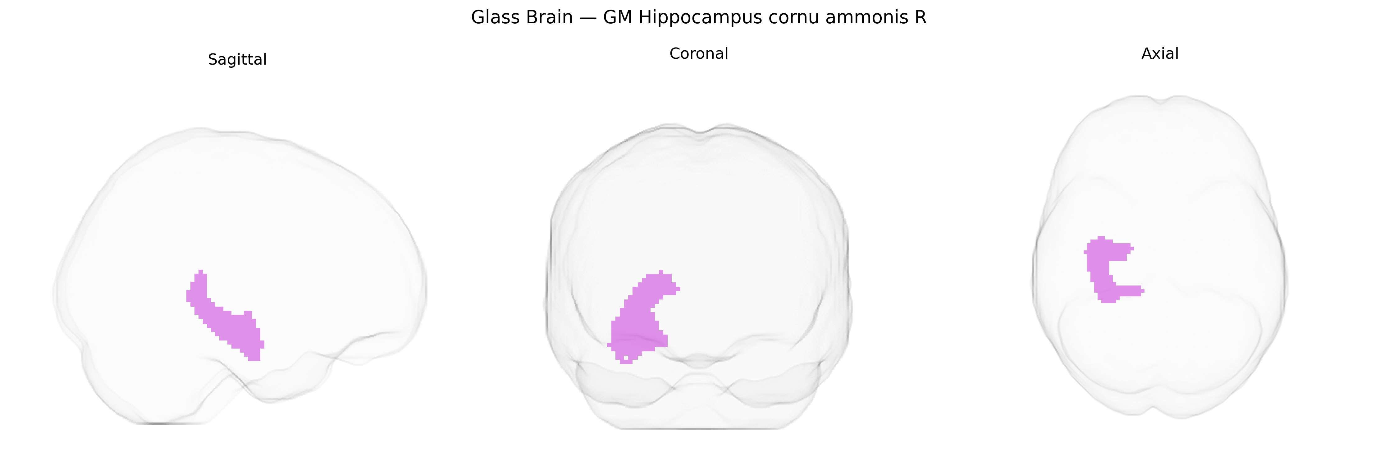
The bilateral GM Hippocampus cornu ammonis R, as defined in the Juelich maxprob thr25 2mm Atlas, corresponds primarily to the cornu ammonis (CA) fields of the right hippocampus, a key allocortical structure within the medial temporal lobe that is critical for episodic memory formation, spatial navigation, and consolidation of short-term into long-term memories. Histologically, this region comprises distinct CA subfields (e.g., CA1–CA4), characterized by densely packed pyramidal neurons arranged in a curved laminar architecture that interfaces with the dentate gyrus and subiculum, forming part of the hippocampal trisynaptic circuit. Functionally, the cornu ammonis participates in pattern separation and completion, contextual encoding, and retrieval processes, and is highly sensitive to hypoxia, neurodegeneration, and stress-related glucocorticoid effects, as seen in conditions such as Alzheimer’s disease, temporal lobe epilepsy, and depression. A closely related and encompassing structure is the Hippocampus.
The bilateral gray matter hippocampal cornu ammonis (CA) subfields from the Juelich maxprob thr25 2 mm atlas correspond to core hippocampal circuitry that has been repeatedly implicated in genetic studies of brain structure and neuropsychiatric disease. Twin and SNP-based heritability analyses show substantial heritability of hippocampal and CA subfield volumes, with genome-wide association studies (GWAS) identifying variants in and around genes such as HMGA2, MSRB3, DPP4, WIF1, ASTN2, and FOXO3, as well as loci in the major histocompatibility complex (MHC), as contributors to individual differences in hippocampal morphology. Large-scale imaging genetics consortia (e.g., ENIGMA, UK Biobank analyses) report that CA-specific volume variation is influenced by polygenic architectures overlapping with those for Alzheimer’s disease, schizophrenia, major depressive disorder, bipolar disorder, and epilepsy, including risk loci in or near APOE, BIN1, SORL1, and other Alzheimer’s-associated genes that are enriched for effects on hippocampal degeneration. Polygenic risk scores for Alzheimer’s disease and schizophrenia show associations with reduced hippocampal and CA volumes, while genetic liability for depression and anxiety disorders correlates with CA subfield alterations. Additionally, GWAS of cognitive traits and educational attainment have identified genetic variants that simultaneously affect hippocampal volume and memory performance, implying shared genetic pathways in neurodevelopment, synaptic plasticity, and neurodegeneration that converge on the cornu ammonis fields.
Overview generated by GPT-4o (2026).
Region ID: 18
Hemisphere: bilateral
Atlas: Juelich maxprob thr25 2mm
Full Quality Version: Download MP4
Full Quality Version: Download MP4
Wali Sidiqyar*, Gaurav Rudravaram*, Elyssa M. McMaster, Trent M. Schwartz, Adam M. Saunders, Kurt G. Schilling, Bennett A. Landman "Introducing SPINS: A Shared Public Visualization Library of Neuroanatomical Structures." Medical Imaging with Deep Learning- short paper
This resource is licensed under CC0 1.0 Universal (Public Domain).