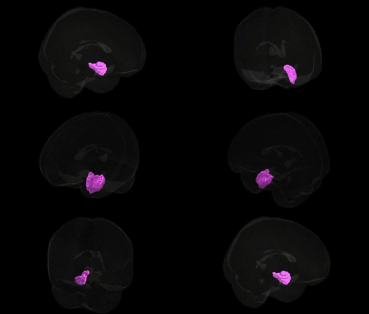
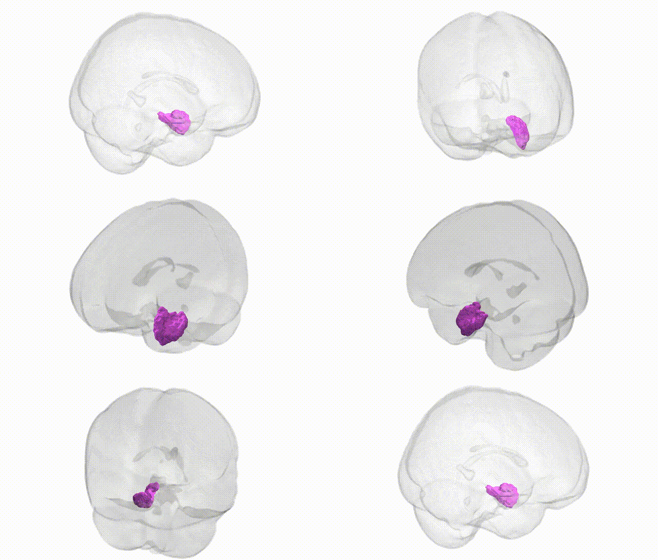
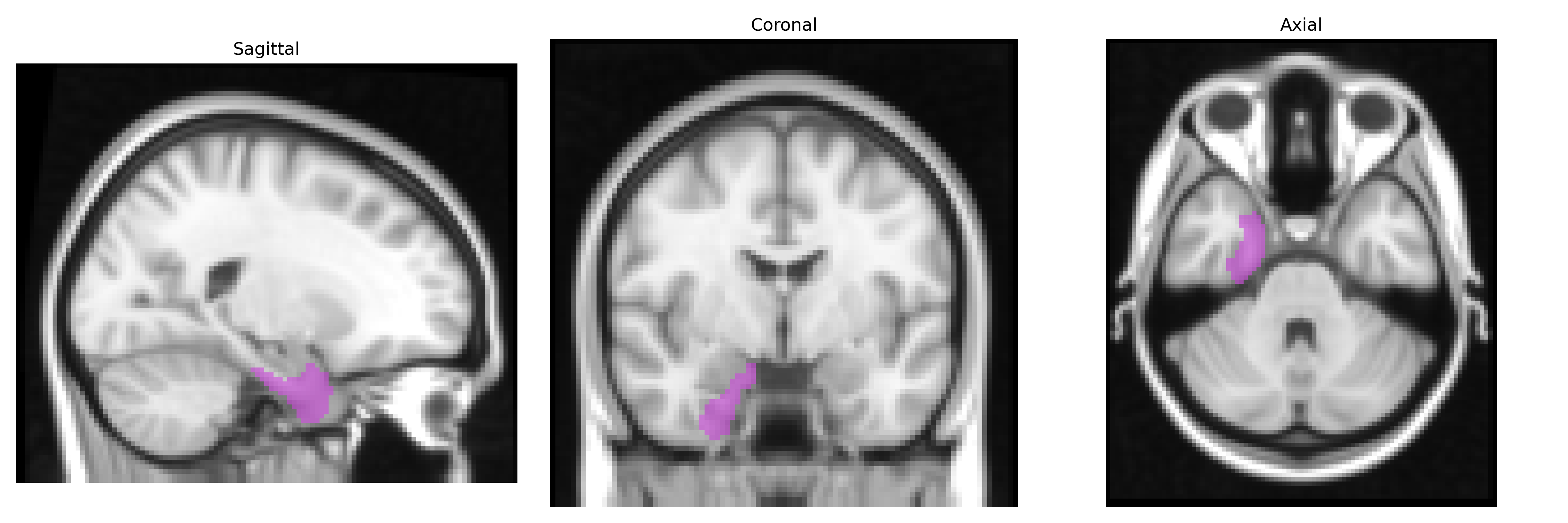
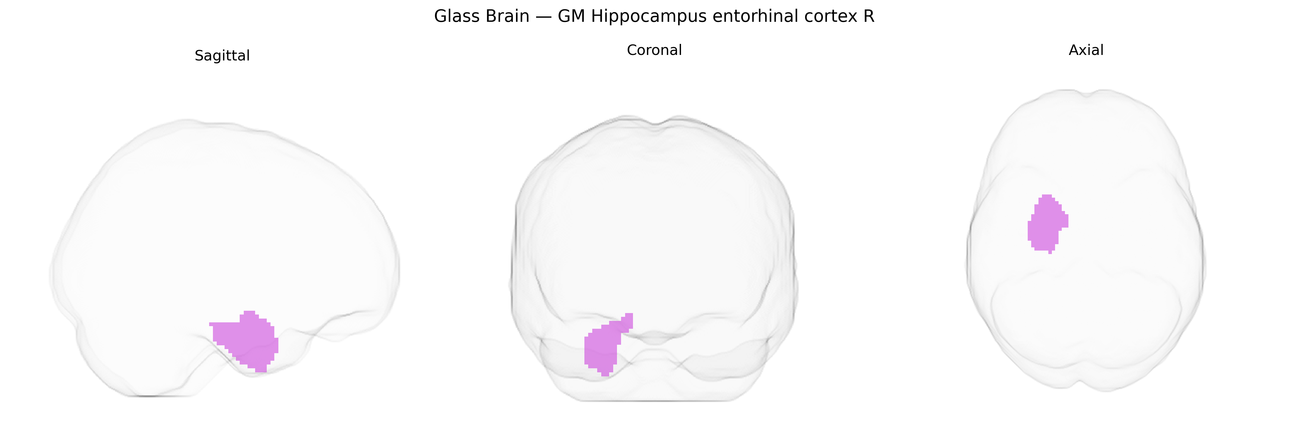
The bilateral gray matter (GM) Hippocampus–Entorhinal Cortex R region in the Juelich maxprob thr25 2mm atlas corresponds to a right-lateralized medial temporal lobe territory encompassing the posterior entorhinal cortex and adjacent hippocampal formation, including portions of the cornu ammonis and subiculum. This region plays a central role in episodic and spatial memory, serving as a hub for the convergence of multimodal sensory information and its relay into the hippocampal circuitry, as well as in the consolidation and retrieval of long-term memories. Cytoarchitectonically, it is characterized by a layered allocortical organization transitioning from the six-layered neocortex of the temporal lobe to the three-layered hippocampal allocortex, with rich reciprocal connections to the neocortex, amygdala, and other limbic structures. There is no single direct Wikipedia article for the combined “Hippocampus–Entorhinal Cortex R” label; related entries include the Hippocampus and the Entorhinal cortex.
The bilateral gray matter hippocampus–entorhinal cortex (right) region in the Juelich maxprob thr25 2mm atlas corresponds to a medial temporal lobe hub strongly implicated in genetic studies of cognition, neurodegeneration, and psychiatric illness. GWAS of hippocampal and entorhinal cortical volume and thickness have consistently identified common variants in genes such as APOE (particularly ε4), CLU, CR1, BIN1, ABCA7, SORL1, and PICALM, many of which are also major risk loci for Alzheimer’s disease and influence atrophy, amyloid/tau burden, and age-related decline in this region. Variants in MAPT, GRIN2B, TOMM40, and genes involved in synaptic plasticity and neurogenesis (e.g., BDNF, especially Val66Met) have been associated with entorhinal/hippocampal structure and function, memory performance, and susceptibility to temporal lobe epilepsy. Polygenic risk scores for Alzheimer’s disease, schizophrenia, and major depressive disorder show associations with reduced hippocampal and entorhinal volumes and altered connectivity, linking widespread risk variants to structural changes in this area. Additional GWAS have related this region’s morphology to educational attainment, general cognitive ability, and neuroticism, suggesting that genetic influences on memory-related circuitry contribute to both disease risk and normal variability in personality and cognition.
Overview generated by GPT-4o (2026).
Region ID: 20
Hemisphere: bilateral
Atlas: Juelich maxprob thr25 2mm
Full Quality Version: Download MP4
Full Quality Version: Download MP4
Wali Sidiqyar*, Gaurav Rudravaram*, Elyssa M. McMaster, Trent M. Schwartz, Adam M. Saunders, Kurt G. Schilling, Bennett A. Landman "Introducing SPINS: A Shared Public Visualization Library of Neuroanatomical Structures." Medical Imaging with Deep Learning- short paper
This resource is licensed under CC0 1.0 Universal (Public Domain).