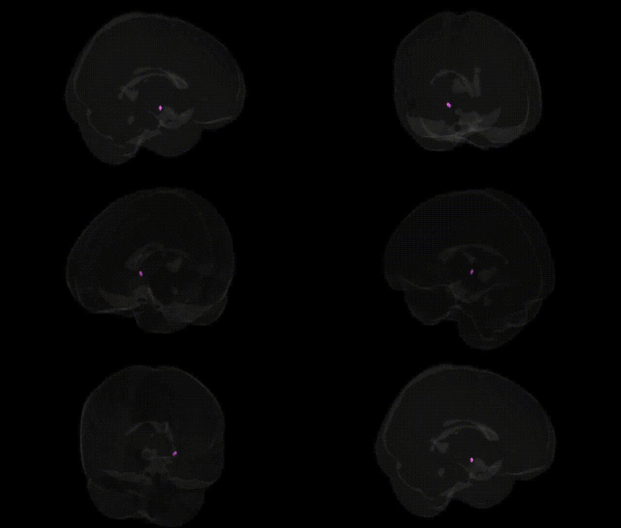
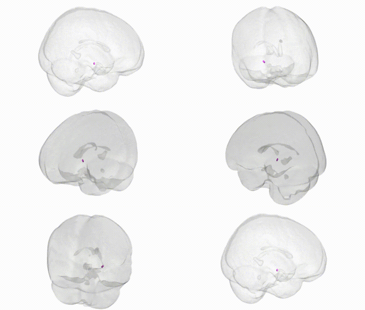
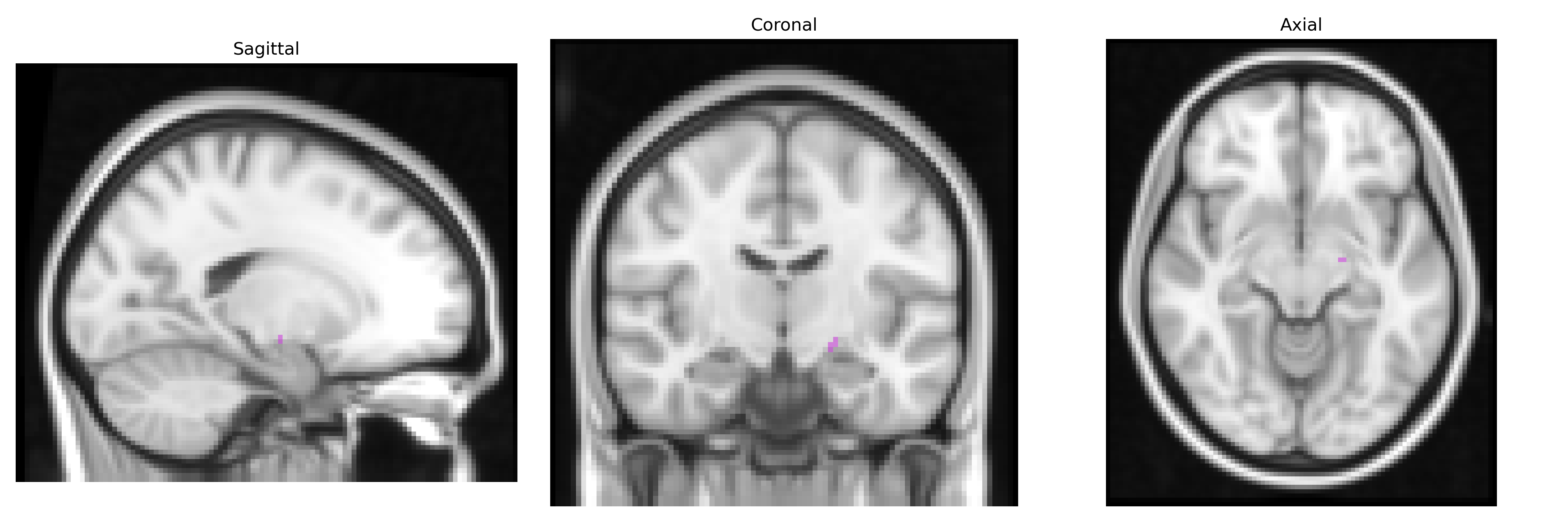
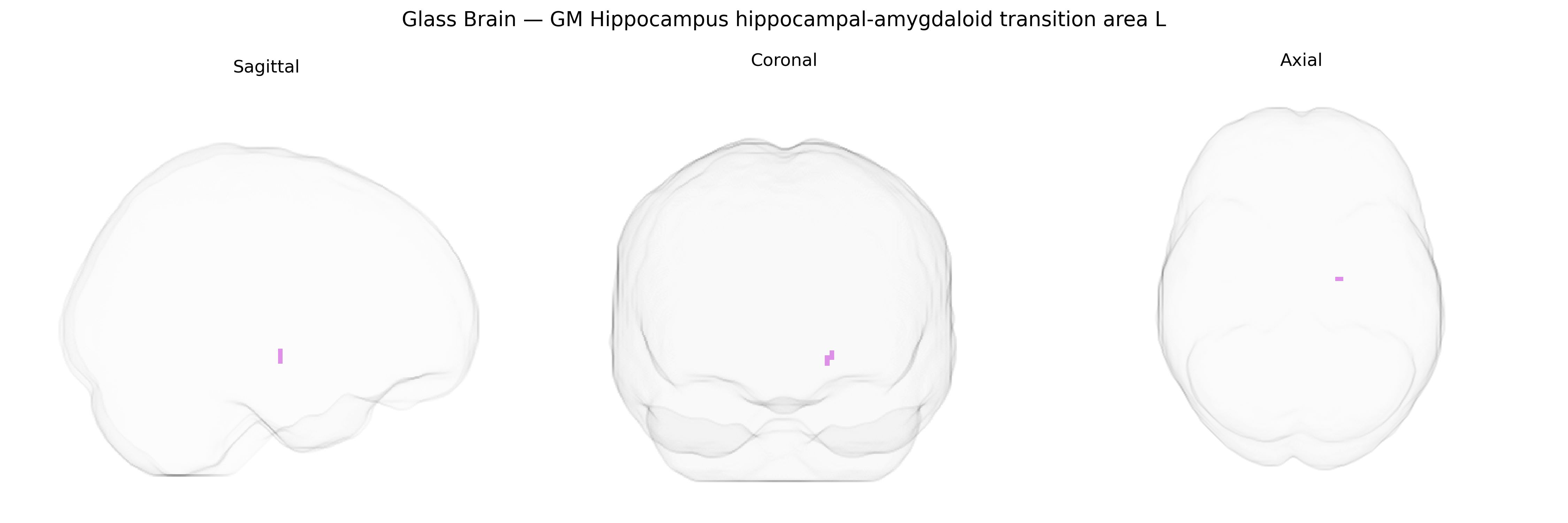
The bilateral GM Hippocampus hippocampal-amygdaloid transition area L, as defined in the Juelich maxprob thr25 2mm Atlas, corresponds to a gray matter region located at the junction between the hippocampus and amygdala within the medial temporal lobe. This transitional zone integrates features of both hippocampal and amygdaloid cytoarchitecture and is implicated in the interface between declarative memory processes and emotional evaluation. It receives multimodal sensory input and contributes to the modulation of memory consolidation by affective salience, linking contextual information encoded by the hippocampus with valence and threat-related processing in the amygdala. While there is no direct Wikipedia entry for this specific transition area, it is closely related to the Hippocampus.
The hippocampal–amygdaloid transition area (HATA), a medial temporal lobe structure lying between the hippocampus and amygdala, has been implicated in several genetic and genome-wide association studies through its contribution to hippocampal and amygdaloid volume, connectivity, and function rather than via HATA-specific loci. Variants in genes related to neurodevelopment and synaptic plasticity—notably BDNF, SLC6A4, COMT, and APOE—have been associated with structural and functional alterations in adjacent hippocampal and amygdala subregions that encompass or closely border the HATA, affecting memory, emotional regulation, and stress responsivity. Large GWAS of subcortical brain volumes (e.g., ENIGMA and UK Biobank consortia) have identified polygenic influences on hippocampal and amygdala morphology, implicating loci near genes involved in neuronal growth, axon guidance, and glutamatergic signaling (such as MSRB3, DPP4, HMGA2, HRK, and others), and these effects are likely to extend into transition zones like HATA given current parcellation resolutions. Clinically, genetic risk for psychiatric and neurodegenerative disorders—major depressive disorder, schizophrenia, anxiety disorders, post-traumatic stress disorder, and Alzheimer’s disease—has been linked to altered hippocampal–amygdala structure and connectivity, with polygenic risk scores and disorder-associated loci (for example, in CACNA1C, C4, and APOE) correlating with volumetric or functional changes that include the broader medial temporal circuitry containing HATA; however, current atlases and GWAS do not yet provide robust, HATA-specific genetic associations distinct from those of the hippocampus and amygdala as a whole.
Overview generated by GPT-4o (2026).
Region ID: 23
Hemisphere: bilateral
Atlas: Juelich maxprob thr25 2mm
Full Quality Version: Download MP4
Full Quality Version: Download MP4
Wali Sidiqyar*, Gaurav Rudravaram*, Elyssa M. McMaster, Trent M. Schwartz, Adam M. Saunders, Kurt G. Schilling, Bennett A. Landman "Introducing SPINS: A Shared Public Visualization Library of Neuroanatomical Structures." Medical Imaging with Deep Learning- short paper
This resource is licensed under CC0 1.0 Universal (Public Domain).