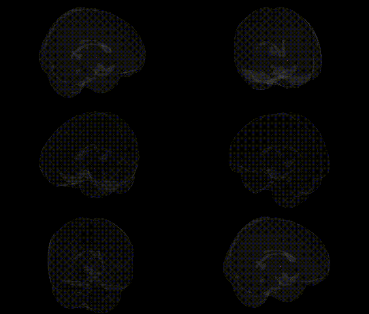
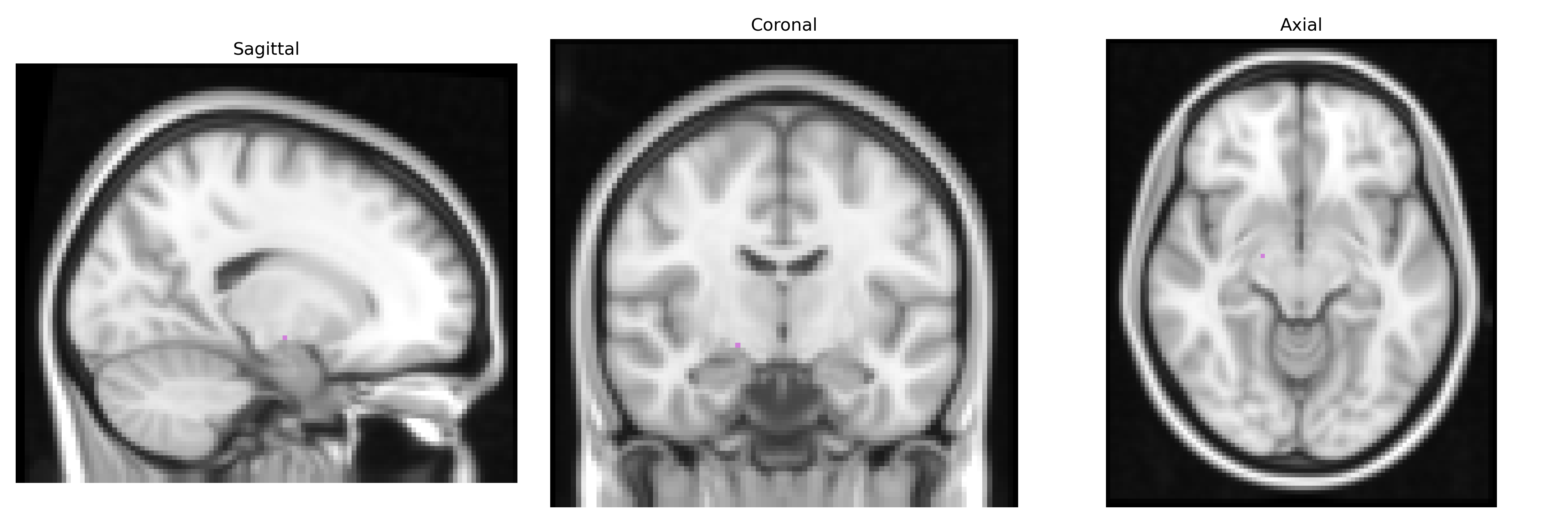
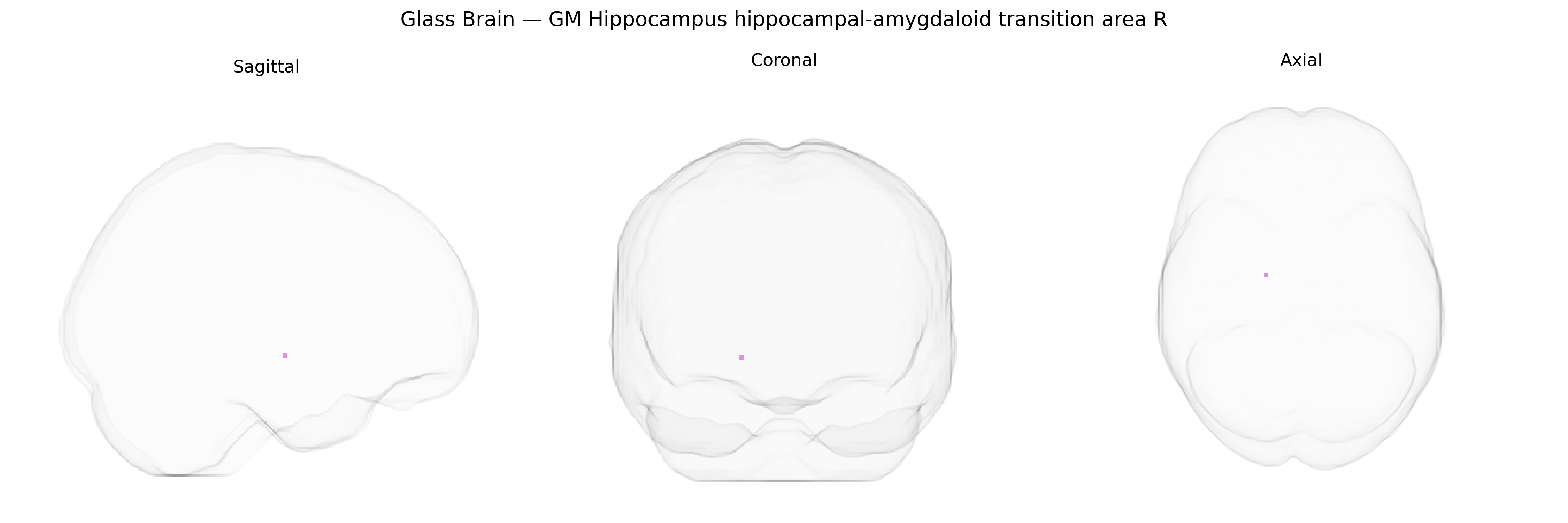
The bilateral GM Hippocampus hippocampal-amygdaloid transition area R, as defined in the Juelich maxprob thr25 2mm Atlas, corresponds to a gray matter region at the interface between the hippocampus and the amygdala within the medial temporal lobe. This transition zone encompasses portions of the hippocampal formation and adjacent cortices that contribute to integrative processing of mnemonic and emotional information, serving as a structural and functional bridge between episodic memory systems and limbic affective networks. Cytoarchitectonically, it includes transitional fields characterized by mixed features of hippocampal and amygdaloid tissue, and functionally it participates in context-dependent emotional memory, fear-related learning, and regulation of stress responses. There is no direct link for the hippocampal-amygdaloid transition area; a related structure is the Hippocampus.
The hippocampal–amygdaloid transition area (HATA), a subregion at the interface of the hippocampus and amygdala as defined in the Juelich maxprob thr25 2 mm atlas, has been implicated in several genetically influenced neuropsychiatric and neurodevelopmental traits through broader hippocampal and amygdalar GWAS and imaging–genetics studies, although direct, high-resolution GWAS focusing specifically on the bilateral GM HATA are limited. Large-scale brain MRI GWAS (e.g., ENIGMA, UK Biobank) have identified common variants in genes such as SLC39A8, DPP4, ASTN2, WNT3, HMGA2, and others that influence hippocampal and amygdalar volumes, with some loci showing pleiotropic associations with cognitive performance, educational attainment, and general brain morphometry. Genetic risk for major depressive disorder, schizophrenia, bipolar disorder, post-traumatic stress disorder, and Alzheimer’s disease (including APOE ε4 and CLU, CR1, PICALM, BIN1 variants) has been associated with structural and functional alterations in hippocampal–amygdalar circuits, including transition zones involved in emotion–memory integration, fear learning, and stress reactivity; polygenic risk scores for these disorders often predict reduced hippocampal volumes and altered amygdala connectivity. Variants in stress-related and neuroplasticity genes such as BDNF (e.g., Val66Met), FKBP5, and genes in the HPA axis and glutamatergic pathways have been linked to differences in hippocampal–amygdaloid structure and function, with downstream influence on anxiety traits, neuroticism, and susceptibility to trauma-related symptoms. Although the Juelich HATA region itself is not yet a primary target in atlas-specific GWAS, converging evidence from fine-grained imaging–genetics work suggests that genetic architectures underlying mood and anxiety disorders, memory performance, and neurodegeneration exert effects on this transition area as part of a broader medial temporal lobe circuit.
Overview generated by GPT-4o (2026).
Region ID: 24
Hemisphere: bilateral
Atlas: Juelich maxprob thr25 2mm
Full Quality Version: Download MP4
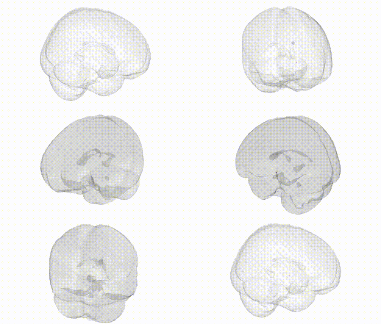
Full Quality Version: Download MP4
Wali Sidiqyar*, Gaurav Rudravaram*, Elyssa M. McMaster, Trent M. Schwartz, Adam M. Saunders, Kurt G. Schilling, Bennett A. Landman "Introducing SPINS: A Shared Public Visualization Library of Neuroanatomical Structures." Medical Imaging with Deep Learning- short paper
This resource is licensed under CC0 1.0 Universal (Public Domain).