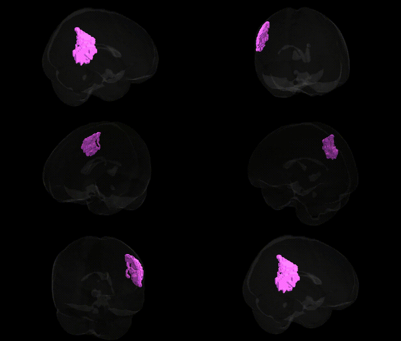
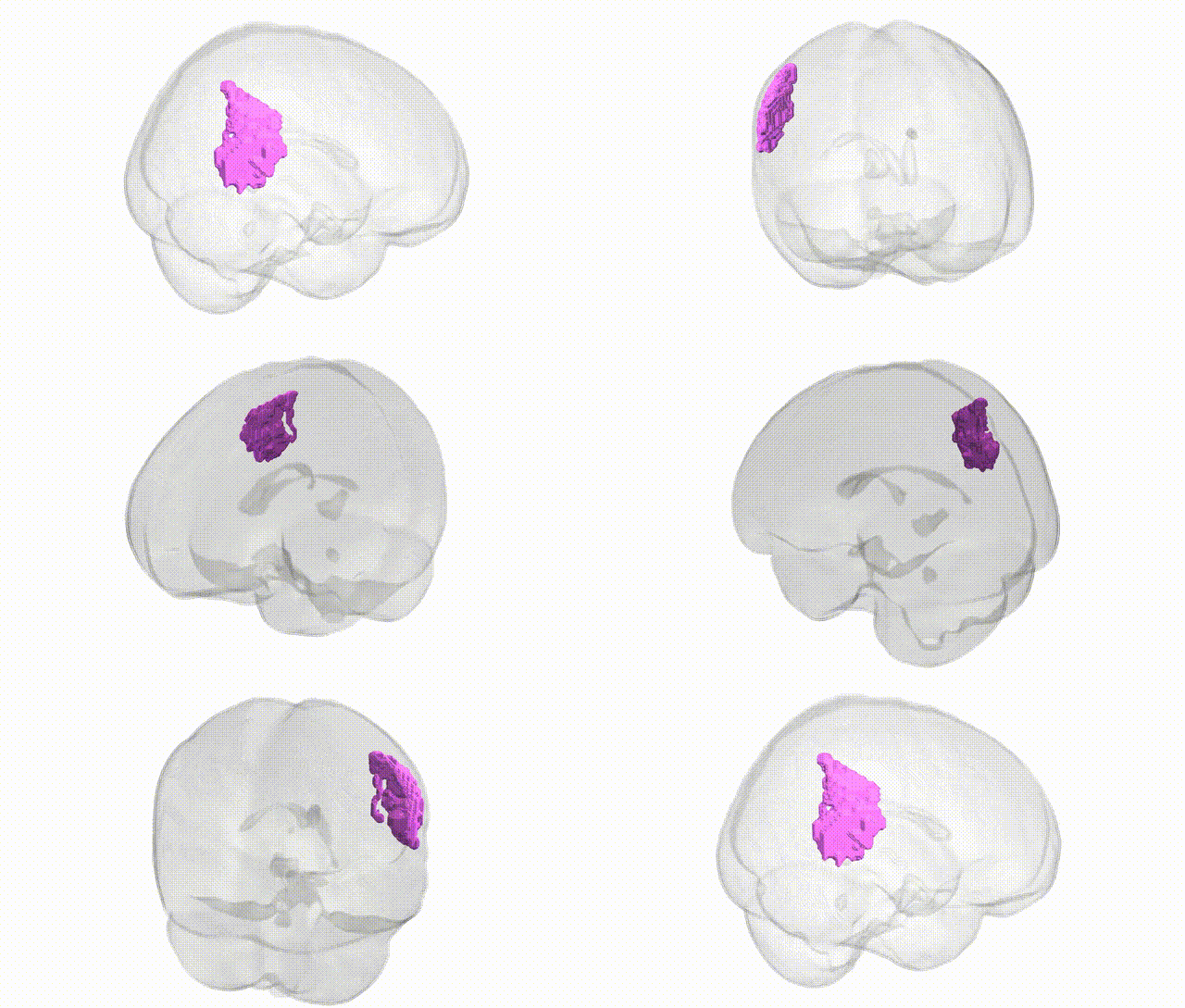
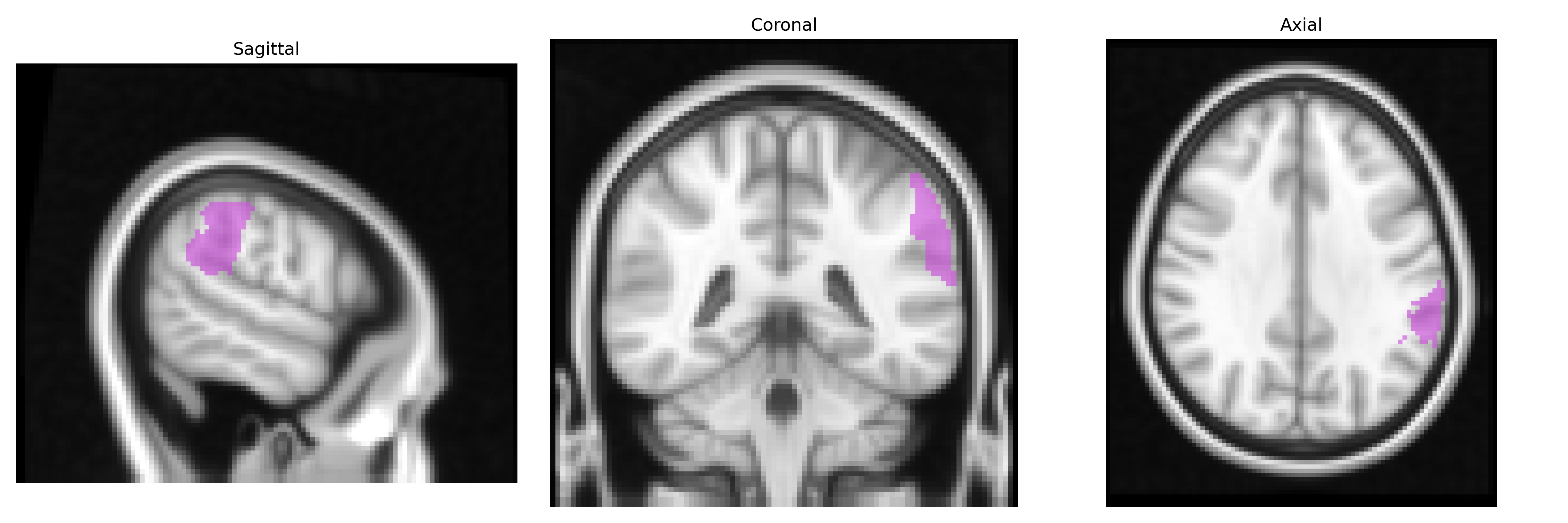
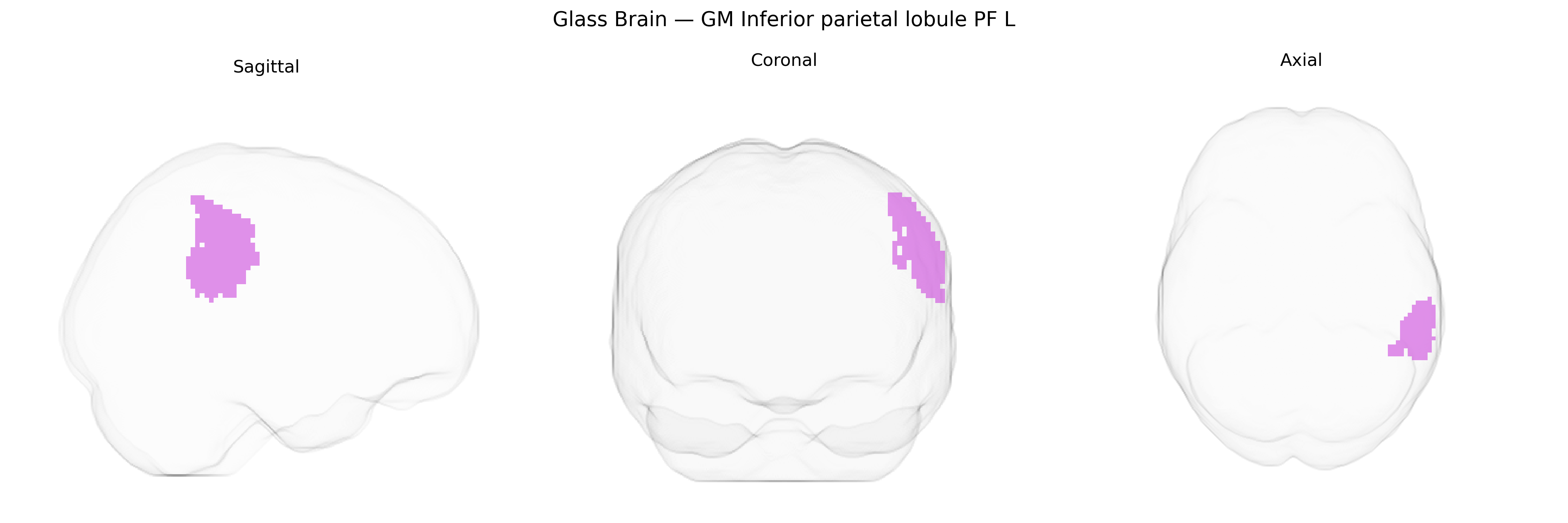
The bilateral GM Inferior parietal lobule PF L (Juelich maxprob thr25 2mm Atlas) refers to a cytoarchitectonically defined subregion of the inferior parietal lobule (IPL) located in the lateral parietal cortex, encompassing portions of the supramarginal and angular gyri. Region PF is part of the inferior parietal cortex associated with multimodal integration of visual, auditory, and somatosensory information, and contributes to higher-order cognitive functions such as attention, language processing, action understanding, and social cognition. It participates in the dorsal attention and frontoparietal control networks and is implicated in processes like spatial awareness, gesture recognition, and aspects of speech perception. There is no direct Wikipedia article for “Inferior parietal lobule PF,” but it is a subregion of the Inferior parietal lobule.
The bilateral GM Inferior parietal lobule PF (left) from the Juelich maxprob thr25 2mm atlas roughly corresponds to parts of the inferior parietal cortex involved in multimodal integration, language, attention, and social cognition, and genetic associations are typically reported at the level of broader parietal or inferior parietal regions rather than the PF subfield specifically. Large-scale brain MRI GWAS (e.g., ENIGMA and UK Biobank–based studies) have identified multiple loci influencing cortical surface area and thickness in inferior parietal and adjacent supramarginal/angular gyri, including variants near genes such as HMGA2, IGF1, and microtubule- or synapse-related genes, though parcellations vary and do not always isolate PF. Parietal and inferior parietal morphometry has been implicated in Alzheimer’s disease risk (with GWAS loci in APOE, CLU, BIN1, and others showing downstream effects on parietal atrophy), schizophrenia and psychosis-related polygenic risk (where higher polygenic risk scores correlate with structural and connectivity alterations in inferior parietal regions), and neurodevelopmental conditions such as autism spectrum disorder and ADHD, in which genetic risk variants affecting synaptic function and neuronal development (e.g., in genes like CNTNAP2, SHANK family, and calcium-channel genes) show associations with altered parietal structure or activation. GWAS of cognitive traits, particularly general intelligence and educational attainment, have also linked polygenic scores to variation in inferior parietal cortical thickness and surface area, consistent with this region’s role in numerical and language-related processing. However, no robust, consistently replicated GWAS has yet pinpointed variants that selectively and uniquely target the PF subregion of the inferior parietal lobule, so current genetic associations remain indirect, inferred from studies of larger parietal territories and related cognitive or neuropsychiatric phenotypes.
Overview generated by GPT-4o (2026).
Region ID: 27
Hemisphere: bilateral
Atlas: Juelich maxprob thr25 2mm
Full Quality Version: Download MP4
Full Quality Version: Download MP4
Wali Sidiqyar*, Gaurav Rudravaram*, Elyssa M. McMaster, Trent M. Schwartz, Adam M. Saunders, Kurt G. Schilling, Bennett A. Landman "Introducing SPINS: A Shared Public Visualization Library of Neuroanatomical Structures." Medical Imaging with Deep Learning- short paper
This resource is licensed under CC0 1.0 Universal (Public Domain).